Előadást letölteni
Az előadás letöltése folymat van. Kérjük, várjon

1
Antigénre nem specifikus immunszuppresszió:
Nem kívánt immunválasz kiküszöbölésére: - Allergia - Autoimmun betegségek - Szervátültetés: kilökődés, GVH Antigén specifikus immunszuppresszió – szelektív tolerancia - cél Antigénre nem specifikus immunszuppresszió: Kortikoszteroidok: gyulladási reakciók gátlása - immunválasz gátlása Mechanizmus: glukokortikoszteroid - hormon receptorokon keresztül Természetben előforduló 21 C atomos szteroid hormon: kortikoszteroid- > a koleszterin metabolizmus terméke 90 % fehérjéhez kötött in vivo –kortikoszteroid kötő globulinhoz kötve >>5-8% albumin –hoz; lebomlás ellen Lebontás helye: máj, 4-5 kötés, keton csoport hidrolízise, konjugálódás: glukuronid, szulfát, 95%- át vese kiválasztja 1948: hidrokortizon (Reumatoid artitisz)
")
3
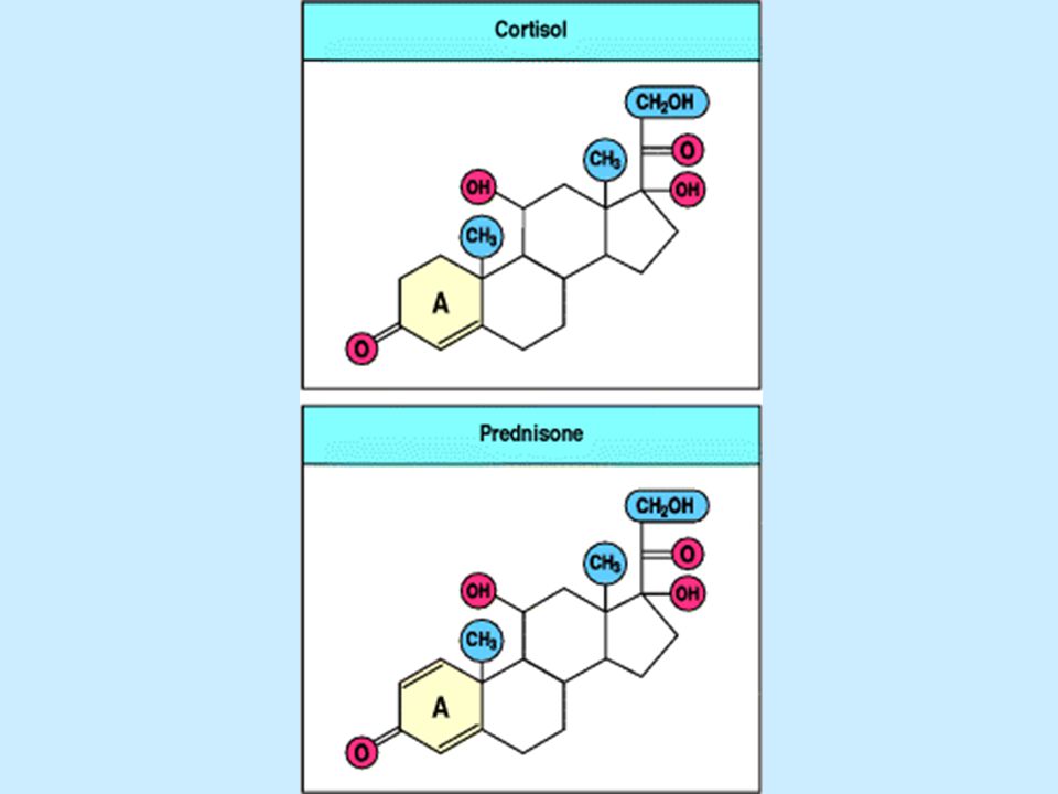
Szerkezete: kortizol prednizolon Kortizon Prednizon
11C-OH - aktív, =O – inaktív (in vivo átalakulás) szintetikus termékek: CH2OH C=O CH2OH C=O OH OH O kortizol prednizolon Kortizon Prednizon (4x hatásosabb) OH O CH2OH C=O O O The structure of the anti-inflammatory corticosteroid drug prednisone. Prednisone is a synthetic analogue of the natural adrenocorticosteroid cortisol. Introduction of the 1,2 double bond into the A ring increases anti-inflammatory potency approximately fourfold compared with cortisol, without modifying the sodium-retaining activity of the compound CH2OH C=O O O
 szintetikus termékek: CH2OH. C=O. CH2OH. C=O. OH. OH. O. kortizol. prednizolon. Kortizon. Prednizon. (4x hatásosabb) OH. O. CH2OH. C=O. O. O. The structure of the anti-inflammatory corticosteroid drug prednisone. Prednisone is a synthetic analogue of the natural adrenocorticosteroid cortisol. Introduction of the 1,2 double bond into the A ring increases anti-inflammatory potency approximately fourfold compared with cortisol, without modifying the sodium-retaining activity of the compound. CH2OH. C=O. O. O.")
4
Szteroid hormonok hatásmechanizmusa
sejtmembrán szteroid citoplazma szteroid receptor HSP-90 citoplazma sejtmag szabályozó gén elem sejtmag transzkripció mRNS transzláció fehérje

5
Hatás: leukociták cirkulációja neutrofil granulociták száma átmenetileg nő ( 6 h) más sejtek száma csökken (limfociták, eozonofil, bazofil, monocita) sejtfunkció megváltoztatása : enzim felszabadulás-nem lizoszomális citokin felszabadulás: IL1, IL-2, TNF, IL-6 limfociták aktiválhatósága, APC funkció fagocitózis gyulladás elleni hatás: gyulladás kialakulásában szerepet játszó sejtek funkciója metabolikus hatás: lebomlást elősegíti, lipid, fehérje, szénhidrát, nukleinsav toxicitás A gének 1%-ának expresszióját szabályozhatja
 sejtfunkció megváltoztatása : enzim felszabadulás-nem lizoszomális citokin felszabadulás: IL1, IL-2, TNF, IL-6 limfociták aktiválhatósága, APC funkció fagocitózis gyulladás elleni hatás: gyulladás kialakulásában szerepet játszó sejtek. funkciója. metabolikus hatás: lebomlást elősegíti, lipid, fehérje, szénhidrát, nukleinsav toxicitás. A gének 1%-ának expresszióját szabályozhatja.")
6
A glukokortikoidok hatása a sejtszámra
4,000- 2,000- 400- 300- 100- 10,000- Neutrofil granulociták Limfociták Eozinofilok Monocyták Bazofilok Sejt/mm3 6 h h h A változás átmeneti. Neutrofil: fokozott termelés a csontvelőben, + csökkent migráció Limfocita: T> B számuk csökken, CD4> CD8 Monocitopénia: gyulladásgátló hatás, baktericid aktivitás, fagocitózis, migráció , FCR C3R szám csökken

7
A kortikoszteroid terápia gyulladásgátló hatása Aktivitás Effektus
Számos gén aktivitását regulálják Aktivitás Effektus IL-1, TNF, GM-CSF, IL-3, IL-4. IL-5, IL-8 Gyulladás (citokinek által közvetített NOS NO Foszfolipáz A2 Ciklooxigenáz2 Lipokortin Prosztaglandinek, leukotrienek Adhéziós molekulák Csökkent migráció Endonukleázok indukciója Apoptózis indukció (limfociták, eozinofilok)
")
8
X Kortikoszteroidok immunszuppresszív hatásának mechanizmusa
IkBa gén kortikoszteroidok Megnövekedett transzkripció és fehérjeszintézis Citokin gén X NF-kB IkBa transzkripció

9
Nem steroid természetű gyulladásgátlók
Aszpirin (fűzfa - Hippokratesz) Szintetikus előállítás : XIX sz., ma USA –15x106 kg / év Hatásmechanizmus: ciklooxigenáz gátlás prosztaglandin termelés gátlása enzim aktív hely: szerin acetilálása (irreverzibilis) arachidonsav kötődés gátlása (reverzibilis)
 Szintetikus előállítás : XIX sz., ma USA –15x106 kg / év. Hatásmechanizmus: ciklooxigenáz gátlás prosztaglandin termelés gátlása. enzim aktív hely: szerin acetilálása (irreverzibilis) arachidonsav kötődés gátlása (reverzibilis)")
10
Foszfolipid - sejtmembrán
Hatásmechanizmus: Foszfolipid - sejtmembrán foszfolipáz A2 arachidonsav 15 HEPTE lipoxin ciklooxigenáz lipoxigenáz vazodilatáció, PGH2 5 lipoxigenáz LTB4 antagonizmus + FLAP TBX PGI PGE 5 HPETE vazodilatáció 5lipoxigenáz trombocita LTA2 LTA4 szintáz aggregáció LTC4 szintáz LTB4 LTC4 transzpeptidáz simaizom kontrakció PMN kemotaxis LTD4 érfal permeabilitás PMN aktiválódás dipeptidáz citokin termelés T sejt ” LTE érfal permeabilitás

11
NF-ATn szintézis, [Ca2+]
A ciklosporin (CsA) és az FK506 immunszuppresszív szerek a citoplazmában hatnak. Különböző célmolekulákhoz kötődnek: a ciklofilinhez (CyP), ill. az FK-kötő fehérjéhez (FKBP) A CsA-CyP és a FK506-FKBP komplexek kötődnek a calcineurinhoz, megakadályozva [Ca2+] általi aktivációját, ezáltal gátolják az NF-Atc aktiválódását T sejt aktiválódás NF-ATn szintézis, [Ca2+] A [ Ca2+] aktiválja a calcineurint, az NF-ATc-t aktiváló foszfatázt, Az aktivált NF-ATc bejut a magba és hozzákötődik az NF-ATn-hez, kialakul az aktív transzkripciós faktor, az NF-AT Figure Cyclosporin A and tacrolimus inhibit T-cell activation by interfering with the serine/threonine-specific phosphatase calcineurin. Signaling via T-cell receptor-associated tyrosine kinases leads to the activation and increased synthesis of the transcription factor AP-1 and other partner proteins, as well as increasing the concentration of Ca2+ in the cytoplasm (left panels). The Ca2+ binds to calcineurin and thereby activates it to dephosphorylate the cytoplasmic form of members of the family of nuclear factors of activated T cells (NFATc). Once dephosphorylated, the active NFATc family members migrate to the nucleus to form a complex with AP-1 and other partner proteins; the NFATc:AP-1 complexes can then induce the transcription of genes required for T-cell activation, including the IL-2 gene. When cyclosporin A (CsA) or tacrolimus are present, they form complexes with their immunophilin targets, cyclophilin (CyP) and FK-binding protein (FKBP), respectively (right panels). The complex of cyclophilin with cyclosporin A can bind to calcineurin and block its ability to activate NFATc family members. The complex of tacrolimus with FKBP binds to calcineurin at the same site, also blocking its activity. Génátírás aktiválása Génátírás nem aktiválódik
![NF-ATn szintézis, [Ca2+]](http://slideplayer.hu/slide/2215054/8/images/11/NF-ATn+szint%C3%A9zis%2C+%5BCa2%2B%5D.jpg "A ciklosporin (CsA) és az FK506 immunszuppresszív szerek a citoplazmában hatnak. Különböző célmolekulákhoz kötődnek: a ciklofilinhez (CyP), ill. az FK-kötő fehérjéhez (FKBP) A CsA-CyP és a FK506-FKBP komplexek kötődnek a calcineurinhoz, megakadályozva [Ca2+] általi aktivációját, ezáltal gátolják az NF-Atc aktiválódását. T sejt aktiválódás. NF-ATn szintézis, [Ca2+] A [ Ca2+] aktiválja a calcineurint, az. NF-ATc-t aktiváló foszfatázt, Az aktivált NF-ATc bejut a magba és hozzákötődik az. NF-ATn-hez, kialakul az aktív transzkripciós faktor, az NF-AT. Figure Cyclosporin A and tacrolimus inhibit T-cell activation by interfering with the serine/threonine-specific phosphatase calcineurin. Signaling via T-cell receptor-associated tyrosine kinases leads to the activation and increased synthesis of the transcription factor AP-1 and other partner proteins, as well as increasing the concentration of Ca2+ in the cytoplasm (left panels). The Ca2+ binds to calcineurin and thereby activates it to dephosphorylate the cytoplasmic form of members of the family of nuclear factors of activated T cells (NFATc). Once dephosphorylated, the active NFATc family members migrate to the nucleus to form a complex with AP-1 and other partner proteins; the NFATc:AP-1 complexes can then induce the transcription of genes required for T-cell activation, including the IL-2 gene. When cyclosporin A (CsA) or tacrolimus are present, they form complexes with their immunophilin targets, cyclophilin (CyP) and FK-binding protein (FKBP), respectively (right panels). The complex of cyclophilin with cyclosporin A can bind to calcineurin and block its ability to activate NFATc family members. The complex of tacrolimus with FKBP binds to calcineurin at the same site, also blocking its activity. Génátírás aktiválása Génátírás nem aktiválódik.")
12
A ciklosporin A és a takrolimusz immunológiai hatásai
T sejtek: IL-2, IL-3, IL-4 GMCSF, TNFa CSÖKKENT EXPRESSZIÓ CSÖKKENT PROLIFERÁCIÓ CSÖKKENT CALCIUM FÜGGŐ EXOCITÓZIS CSÖKKENT APOPTÓZIS (AICD) B sejtek: CSÖKKENT PROLIFERÁCIÓ A T SEJTES SEGÍTSÉG HIÁNYA MIATT CSÖKKENT PROLIFERÁCIÓ ANTI-Ig KERESZTKÖTÉSRE CSÖKKENT APOPTÓZIS Granulociták: CSÖKKENT CALCIUM FÜGGŐ EXOCITÓZIS (szerin eszteráz tartalmú granulumok) Rapamycin: FK506-hoz hasonló, CD28 jelátvitelt gátol., nem Ca2+ függő
 B sejtek: CSÖKKENT PROLIFERÁCIÓ A T SEJTES SEGÍTSÉG HIÁNYA MIATT CSÖKKENT PROLIFERÁCIÓ ANTI-Ig KERESZTKÖTÉSRE. CSÖKKENT APOPTÓZIS. Granulociták: CSÖKKENT CALCIUM FÜGGŐ EXOCITÓZIS (szerin eszteráz tartalmú granulumok) Rapamycin: FK506-hoz hasonló, CD28 jelátvitelt gátol., nem Ca2+ függő.")
13
Citotoxikus szerek: Az osztódó sejteket pusztítják el
Az osztódó sejteket pusztítják el -DNS szintézis fázisában ( azatioprin, metotrexát), vagy -minden fázisban (ciklofoszfamid) Specifitás? Sejtciklusra nem specifikus (UV, besugárzás) S-fázis- specifikus (azatioprin, metotrexát)) Sejtciklus-specifikus (ciklofoszfamid, klorambucil) % ellenanyagtermelő sejt 100 - 10 - 1 - 0.1 - dózis 24 órával az antigén előtt adva 24 órával az antigén után adva Különböző immunszuppresszív szerek hatása az ellenanyagtermelő sejtek számára
, vagy. -minden fázisban (ciklofoszfamid) Specifitás Sejtciklusra nem specifikus (UV, besugárzás) S-fázis- specifikus (azatioprin, metotrexát)) Sejtciklus-specifikus (ciklofoszfamid, klorambucil) % ellenanyagtermelő sejt dózis. 24 órával az antigén előtt adva. 24 órával az antigén után adva. Különböző immunszuppresszív szerek hatása az ellenanyagtermelő sejtek számára.")
14
Citotoxikus immunszuppresszív szerek szerkezete és metabolizmusa
Purin bioszintézist gátolja DNS szintézisfázisa The structure and metabolism of the cytotoxic immunosuppressive drugs azathioprine and cyclophosphamide. Azathioprine was developed as a modification of the anti-cancer drug 6-mercaptopurine; by blocking the reactive thiol group, the metabolism of this drug is slowed down. It is slowly converted in vivo to 6-mercaptopurine, which is then metabolized to 6-thio-inosinic acid, which blocks the pathway of purine bio-synthesis. Cyclopho-sphamide was similarly developed as a stable pro-drug, which is activated enzymatically in the body to phosphoramide mustard, a powerful and unstable DNA-alkylating agent. DNS alkilező szer, nem stabil Minden fázisban

15
A ciklofoszfamid hatása a limfocitákra: elsősorban a DNS szálak keresztkötése
B sejtek általában lassabban regenerálódnak a hatás kifejezettebb mint T sejteken

16
Rh negatív terhes anyák megelőző anti-D IgG terápiája:
antigén specifikus immunszuppresszió

17
Az anti-D IgG lehetséges hatásmechanizmusai
a: BCR-FcgRIIb keresztkötés, b: BCR-FcgRIIb keresztkötés K antigénen keresztül, c: az anti-D IgG keresztköti a BCR-t és az FcgRIIb-t, d: D antigén maszkírozása vvt-n, e: fagocitózis az FcgRI vagy FcgRIII-on keresztül

18
Poliklonális ellenanyagok:
Nem ag specifikus immunszuppresszió- immunmoduláció Állatok humán limfocitákkal való immunizálása: anti-timocita szérum, anti-limfocita szérum T sejtes választ gátolja Limfopénia(?) Problémák: standardizáció, nem szelektív a T sejtekre idegen fehérje –immunválsz (szérum betegség) ezért:
 Problémák: standardizáció, nem szelektív a T sejtekre. idegen fehérje –immunválsz (szérum betegség) ezért:")
19
Monoklonális Ellenanyagok:
Homogén Szelektivitás Humanizált ellenanyagok – nincs immunválasz Graft kilökődés elkerülésére, APC és/vagy T sejt receptoraihoz kötődnek- immunszuppresszió Okt3 (CD3) : limfopénia, mechanizmuns nem tisztázott Ujra megjelenő sejtek funkciója gátolt Human anti-egér IgG! termelést vált ki Aktiválhatja a T sejteket ! Citokin felszabadulás, gyulladás – +corticosteroid Immunszuppresszió, limfoproliferatív betgségek megjelenése MHCII, CD4, gátló hatás LFA1, ICAM1 – sikeresen alkalmazták CD28, B7 IL-2R Jelölt ellenanyagok: radioaktiv, toxin etc.
 : limfopénia, mechanizmuns nem tisztázott. Ujra megjelenő sejtek funkciója gátolt. Human anti-egér IgG! termelést vált ki. Aktiválhatja a T sejteket ! Citokin felszabadulás, gyulladás – +corticosteroid. Immunszuppresszió, limfoproliferatív betgségek megjelenése. MHCII, CD4, gátló hatás. LFA1, ICAM1 – sikeresen alkalmazták. CD28, B7. IL-2R. Jelölt ellenanyagok: radioaktiv, toxin etc.")
20
Antigen given orally can lead to protection against autoimmune disease.
Figure Antigen given orally can lead to protection against autoimmune disease. Experimental allergic encephalomyelitis (EAE) is induced in mice by immunization with spinal cord homogenate in complete Freund's adjuvant (upper panels); the disease is mediated by TH1 cells specific for myelin antigens. These cells produce the cytokine IFN-γ (top left photograph, where the brown staining reveals the presence of IFN-γ), but not of TGF-β. These T cells are presumably responsible for the damage that results in paralysis. When mice are first fed with myelin basic protein (MBP), later immunization with spinal cord or MBP fails to induce the disease (lower panels). In these orally tolerized mice, IFN-γ-producing cells are absent (lower left photograph), whereas TGF-β-producing T cells (lower right photograph, brown staining) are found in the brain in place of the autoaggressive TH1 cells and presumably protect the brain from autoimmune attack. Photographs courtesy of S. Khoury, W. Hancock, and H. Weiner.
 is induced in mice by immunization with spinal cord homogenate in complete Freund s adjuvant (upper panels); the disease is mediated by TH1 cells specific for myelin antigens. These cells produce the cytokine IFN-γ (top left photograph, where the brown staining reveals the presence of IFN-γ), but not of TGF-β. These T cells are presumably responsible for the damage that results in paralysis. When mice are first fed with myelin basic protein (MBP), later immunization with spinal cord or MBP fails to induce the disease (lower panels). In these orally tolerized mice, IFN-γ-producing cells are absent (lower left photograph), whereas TGF-β-producing T cells (lower right photograph, brown staining) are found in the brain in place of the autoaggressive TH1 cells and presumably protect the brain from autoimmune attack. Photographs courtesy of S. Khoury, W. Hancock, and H. Weiner.")
21
A tolerancia T sejtekkel átvihető
A tissue graft given together with anti-CD4 antibody can induce specific tolerance A tolerancia T sejtekkel átvihető Figure A tissue graft given together with anti-CD4 antibody can induce specific tolerance. Mice grafted with tissue from a genetically different mouse reject that graft. Having been primed to respond to the antigens in the graft, they then reject a subsequent graft of identical tissue more rapidly (left panels). Mice injected with anti-CD4 antibody alone can recover immune competence when the antibody disappears from the circulation, as shown by a normal primary rejection of graft tissue (center panels). However, when tissue is grafted and anti-CD4 antibody is administered at the same time, the primary rejection response is markedly inhibited (right panels). An identical graft made later in the absence of anti-CD4 antibody is not rejected, showing that the animal has become tolerant to the graft antigen. This tolerance can be transferred with T cells to naive recipients (not shown).
. Mice injected with anti-CD4 antibody alone can recover immune competence when the antibody disappears from the circulation, as shown by a normal primary rejection of graft tissue (center panels). However, when tissue is grafted and anti-CD4 antibody is administered at the same time, the primary rejection response is markedly inhibited (right panels). An identical graft made later in the absence of anti-CD4 antibody is not rejected, showing that the animal has become tolerant to the graft antigen. This tolerance can be transferred with T cells to naive recipients (not shown).")
22
SLE vagy más autoimmune betegség:
Okozati kezelés: peptid – pl. SLE hez kapcsolódó autoantigénnek megfelelő peptidek Tolerancia indukció Patogén ellenenyagtermelés megakadályozása Citokin network szabályozása Jelátviteli folyamatok „ apoptózis alapú terápiák

23
Anti-inflammatory effects of anti-TNF-α therapy in rheumatoid arthritis.
Figure Anti-inflammatory effects of anti-TNF-α therapy in rheumatoid arthritis. The clinical course of 24 patients was followed for 4 weeks after treatment with either a placebo or a monoclonal antibody against TNF-α at a dose of 10 mg kg-1. The antibody therapy was associated with a reduction in both subjective and objective parameters of disease activity (as measured by pain score and swollen-joint count, respectively) and in the systemic inflammatory acute-phase response, measured as a fall in the concentration of the acute-phase reactant C-reactive protein. Data courtesy of R.N. Maini.
 and in the systemic inflammatory acute-phase response, measured as a fall in the concentration of the acute-phase reactant C-reactive protein. Data courtesy of R.N. Maini.")
24
Inputs into the core cell-death machinery.
Szignál-terápia – szelektivitás?? (pl. tirozin kináz gátló szerek) Inputs into the core cell-death machinery. Figure 5 | Inputs into the core cell-death machinery. Some of the signal-transduction inputs into core components of the apoptosis machinery are depicted, with a focus on NF-κB, AKT and p53 (see text for details). AKT, v-Akt murine thymoma viral oncogene homologue; APAF1, apoptotic protease-activating factor 1; ASK1, apoptosis signal-regulating kinase 1; BAD, BCL2- antagonist of cell death; BAX, BCL2-associated X protein; DR, death-inducing receptor; FADD, FAS-associated protein with death domain; FASL, FAS ligand; FKHD, forkhead domain; FLIP, FLICE-inhibitory protein; IAP, inhibitor of apoptosis; IKK, IκB kinase; IκB, inhibitor of NF-κB; NF-κB, nuclear factor-κB ; PARP, poly-ADP ribosylpolymerase; PI3K, phosphatidylinositol-3-kinase; PTEN, phosphatase and tensin homologue.
 Inputs into the core cell-death machinery. Figure 5 | Inputs into the core cell-death machinery. Some of the signal-transduction inputs into core components of the. apoptosis machinery are depicted, with a focus on NF-κB, AKT and p53 (see text for details). AKT, v-Akt murine thymoma viral. oncogene homologue; APAF1, apoptotic protease-activating factor 1; ASK1, apoptosis signal-regulating kinase 1; BAD, BCL2- antagonist of cell death; BAX, BCL2-associated X protein; DR, death-inducing receptor; FADD, FAS-associated protein with death. domain; FASL, FAS ligand; FKHD, forkhead domain; FLIP, FLICE-inhibitory protein; IAP, inhibitor of apoptosis; IKK, IκB kinase; IκB, inhibitor of NF-κB; NF-κB, nuclear factor-κB ; PARP, poly-ADP ribosylpolymerase; PI3K, phosphatidylinositol-3-kinase; PTEN, phosphatase and tensin homologue.")
Hasonló előadás


>")


>")
>")


