Előadást letölteni
Az előadás letöltése folymat van. Kérjük, várjon

1
Jelátviteli mechanizmusokhttp://www.bergen.org/ACADEMY/Bio/AnP/AnP1/AnP1Tri2/FIGS/TRANS/phototransduction.html

2
Jelátvivő molekulák: -Sejtmembránon átjutó - vagy át nem jutó molekulák - kontakt szignáling

3
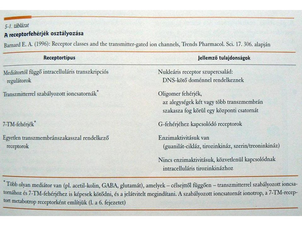
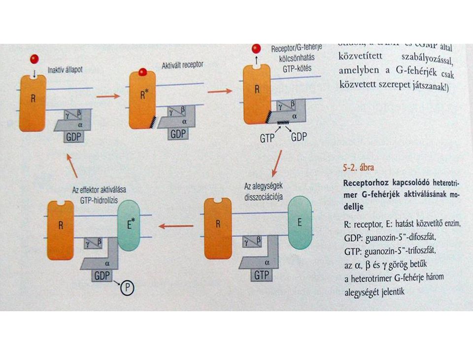
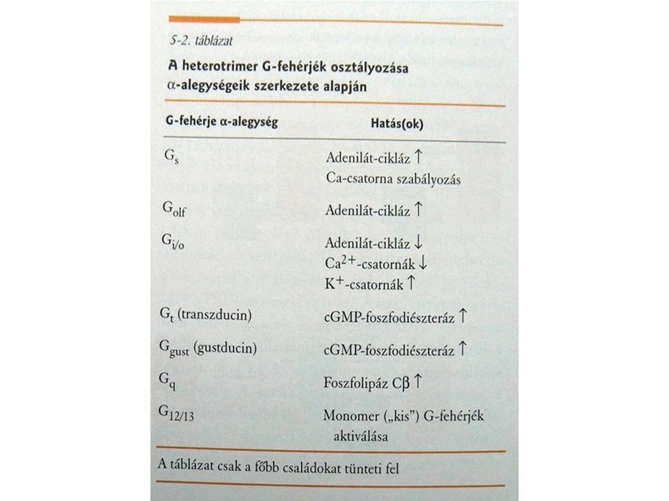
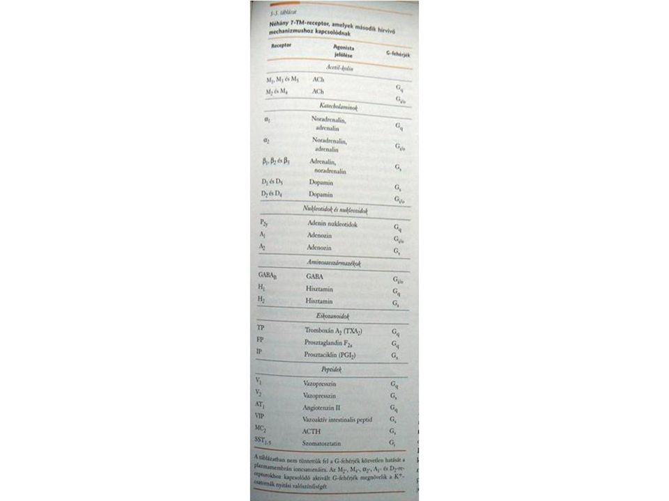
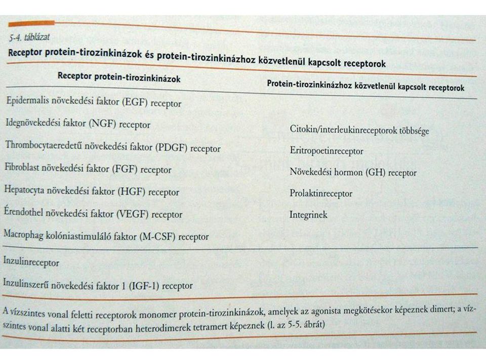
Receptor típusok: -Ion csatornák - Enzimhez direkt módon kapcsolt receptorok - G-proteinez kapcsolt receptorok - Intracelluláris receptorok - Foszforiláció, monomer G-protein

5
Glutamát receptorok Receptorok csoportosítása: Agonisták és antagonisták valamint gén szekvencia alapján http://www.ucl.ac.uk/~smgxt01/frameh.htm?page=glutamat.htm

8
Jel erősítési funkció

10
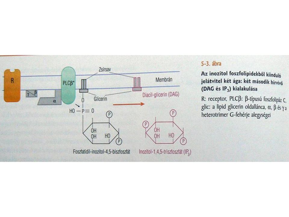
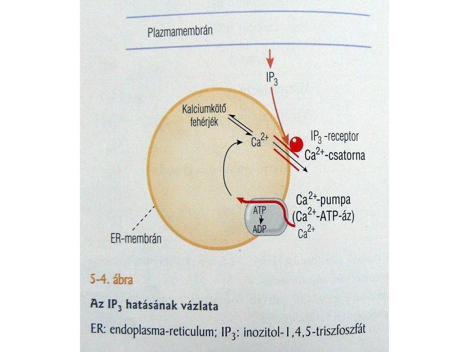
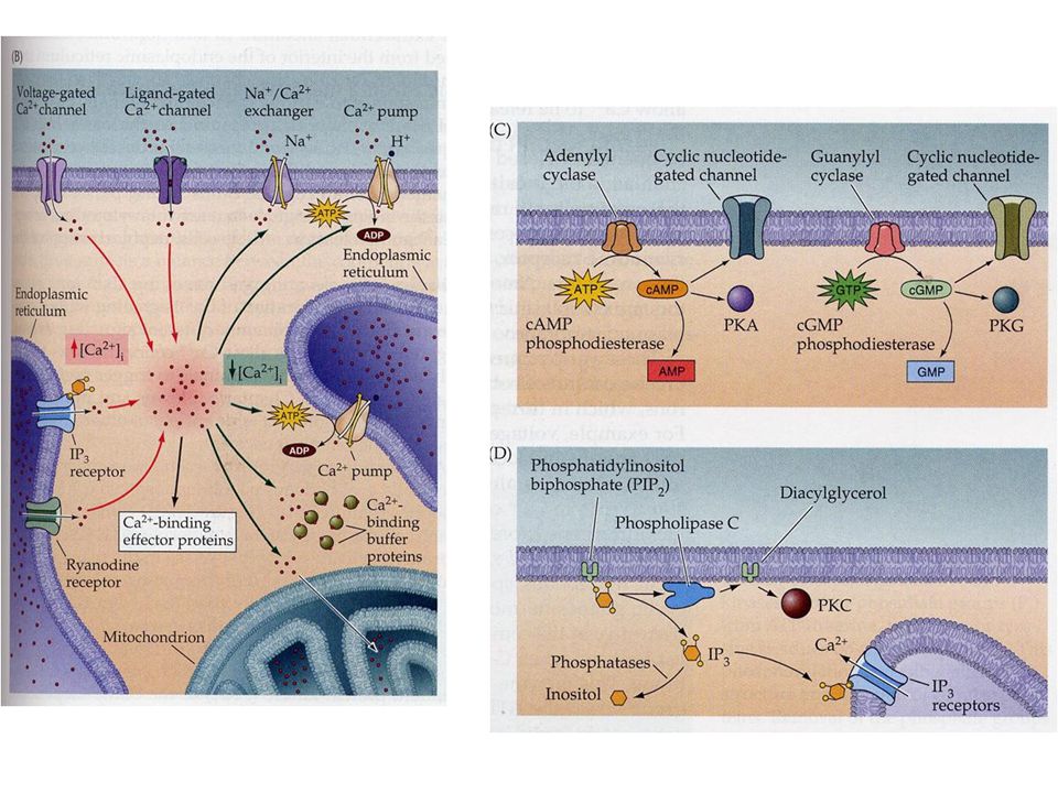
Inositol trifoszfát (IP3) jelátviteli mechanizmus (Intracelluláris kalcium reguláció)
 jelátviteli mechanizmus (Intracelluláris kalcium reguláció)")
13
PIP2: Phosphatidylinositol bisphosphate Másodlagos hírvivők

15
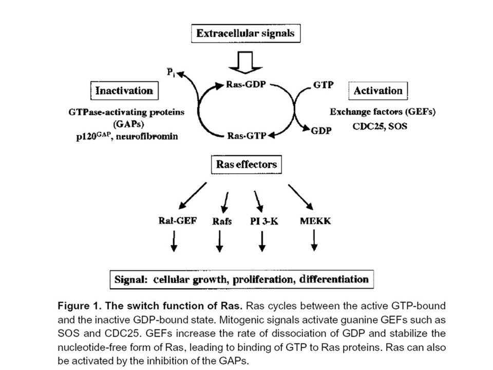
Proteins in the Ras family are very important molecular switches for a wide variety of signal pathways that control such processes as cytoskeletal integrity, proliferation, cell adhesion, apoptosis, and cell migration. Ras and ras related proteins are often deregulated in cancers, leading to increased invasion and metastasis, and decreased apoptosis. The Ras superfamily includes the Ras, Rho, and Rab families.signal pathwaysapoptosis metastasisRhoRab Monomeric G Proteins (small GTPases) homologous to the alpha (α) subunit
 homologous to the alpha (α) subunit.")
17
PKA (cAMP) CaMKII (Ca) PKC (DAG, Ca) Protein tyrosine kinases Mitogen-activated protein kinase /MAPK/ = extracellular signal regulated kinase /ERK/ Phosphates PP1, PP2A PP2B /calcineurin/ Foszforiláción alapuló jelátvitel
 CaMKII (Ca) PKC (DAG, Ca) Protein tyrosine kinases Mitogen-activated protein kinase /MAPK/ = extracellular signal regulated kinase /ERK/ Phosphates PP1, PP2A PP2B /calcineurin/ Foszforiláción alapuló jelátvitel")
21
Regulation of gene expression

22
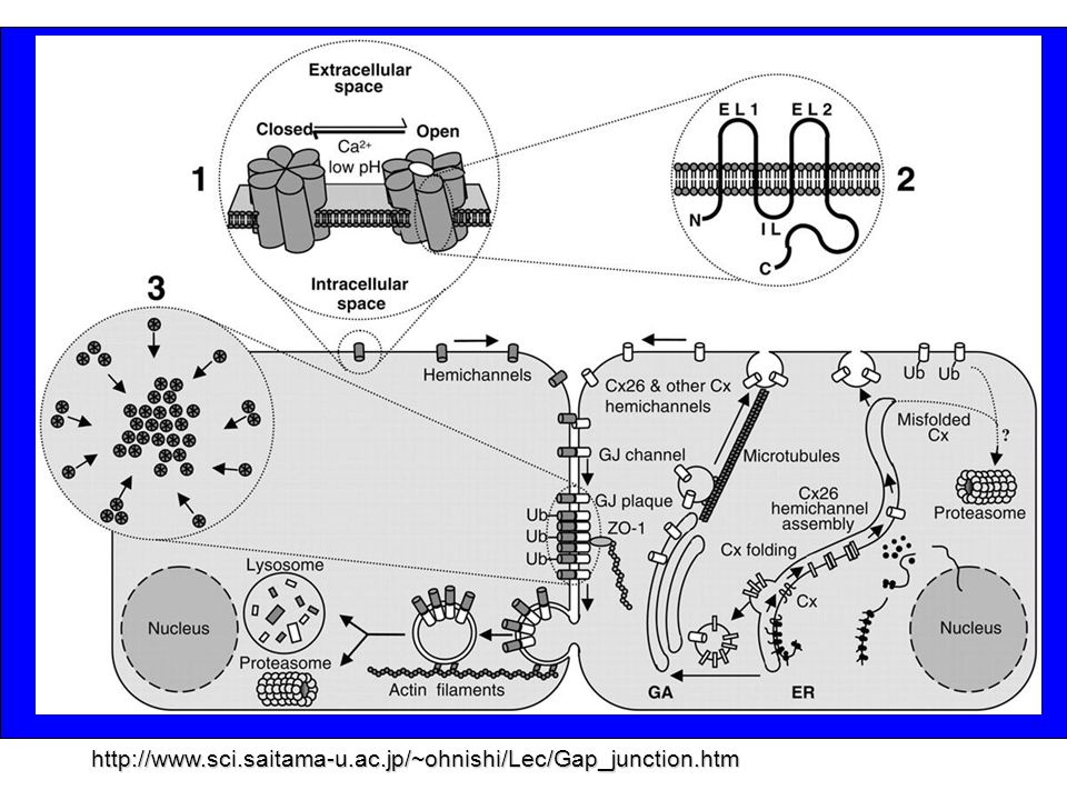
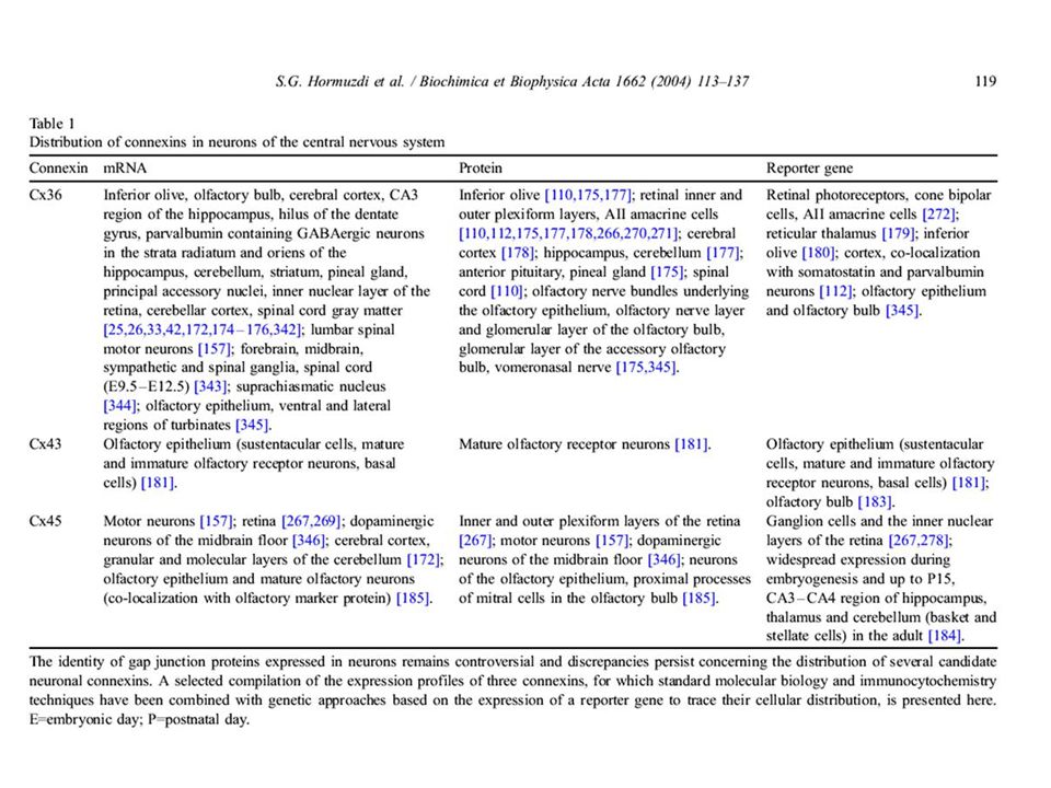
Kémiai szinapszis Neurotranszmitter mediálja a transzmissziót (szinaptikus késés) Szinaptikus rés: 20 – 40 nm Szinaptikus vezikulák, kalciumfüggő transzmitter kibocsátás, pre és posztszinaptikus receptorok, diffúzió, transzmitter bontó enzimek, felvétel http://cla.calpoly.edu/~cslem/Wizdemo/3-ChapterSplash.html http://faculty.etsu.edu/currie/synapse. htm Szinapszis: Specializált struktúra, mely két sejt közötti funkcionális kölcsönhatást biztosítja. Alkotórészei: preszinaptikus terminál, posztszinaptikus célterület és köztük a szinaptikus rés (Biochimica et Biophysica Acta 1662 (2004) 113–137) Elektromos szinapszis (gap junction) Közvetlen kapcsolat a sejtek között csatornákon keresztül (nincs késés, kis molekulák cseréje) 2 nm rés a membránok között Connexinek Kétirányú (de lehet aszimmetrikus)
 113–137) Elektromos szinapszis (gap junction) Közvetlen kapcsolat a sejtek között csatornákon keresztül (nincs késés, kis molekulák cseréje) 2 nm rés a membránok között Connexinek Kétirányú (de lehet aszimmetrikus).")
23
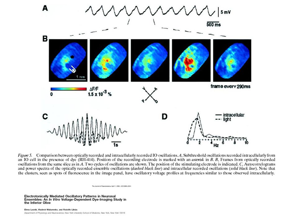
http:// www.colorado.edu/kines/Class/IPHY3430-200/04neuron.html ‘NON-SYNAPTIC’ MECHANISMS IN SEIZURES AND EPILEPTOGENESIS* F. EDWARD DUDEK*, THOMAS YASUMURA and JOHN E. RASH Hogyan tanulmányozzák az elektromos szinapszisokat : Festék átvitel Festék átvitel Elektromos kapcsolat (nincs szinaptikus késés,farmakológia) Elektromos kapcsolat (nincs szinaptikus késés,farmakológia) Transzgén állatokTranszgén állatok Role of Gap Junctions in Synchronized Neuronal Oscillations in the Inferior Olive Elena Leznik and Rodolfo Llina´s http://www.sci.saitama- u.ac.jp/~ohnishi/Lec/Gap_j unction.htm
 Elektromos kapcsolat (nincs szinaptikus késés,farmakológia) Transzgén állatokTranszgén állatok Role of Gap Junctions in Synchronized Neuronal Oscillations in the Inferior Olive Elena Leznik and Rodolfo Llina´s u.ac.jp/~ohnishi/Lec/Gap_j unction.htm.")
25
http://www.sci.saitama-u.ac.jp/~ohnishi/Lec/Gap_junction.htm
26
http://academic.brooklyn.cuny.edu/biology/bio4fv/page/gap-junctions.html Activity-dependent Neuronal Control of Gap-junctional Communication in Astrocytes Nathalie Rouacha, Jacques Glowinskia, and Christian Giaumea Nature. 1993 Mar 25;362(6418):318-24 Magas intracelluláris kalcium (sejt sérülése) zárja a csatornákat
: Magas intracelluláris kalcium (sejt sérülése) zárja a csatornákat.")
27
Figure 1. A model for the interaction of v-Src with Cx43. In this model, the binding of Cx43 to v-Src is dependent initially upon a SH3 domain interaction followed by SH2 domain interactions, which are important for v-Src– induced phosphorylation of Cx43 at the Y265 site and the subsequent phosphorylation at the Y247 site, leading to closure of the Cx43 channel. PXXP denotes the P274–P284 proline-rich sequence of Cx43 that interacts with the SH3 domain of v-Src. For simplicity, gap junction channels are depicted as cylinders. PM denotes the plasma membrane. http://www.crch.org/ProfileLau.htm Szabályozás: Foszforizációval

29
A (kémiai) szinapszis http://www.itg.uiuc.edu/exhibits/gallery/pages/image-51.htm http://www.meddean.luc.edu/lumen/meded/neuro%20/lectures/Transmission.pdf Hogyan tanulmányozzák: Electron mikroszkópElectron mikroszkóp Preszinaptikus stimuláció posztszinaptikus áramot vált ki, amely antagonistákkal gátolhatóPreszinaptikus stimuláció posztszinaptikus áramot vált ki, amely antagonistákkal gátolható Visszafordítási potenciálVisszafordítási potenciál Spontán transzmitter kibocsátás (mEPSPs, mIPSPs)Spontán transzmitter kibocsátás (mEPSPs, mIPSPs) Quantal analízisQuantal analízis
 szinapszis Hogyan tanulmányozzák: Electron mikroszkópElectron mikroszkóp Preszinaptikus stimuláció posztszinaptikus áramot vált ki, amely antagonistákkal gátolhatóPreszinaptikus stimuláció posztszinaptikus áramot vált ki, amely antagonistákkal gátolható Visszafordítási potenciálVisszafordítási potenciál Spontán transzmitter kibocsátás (mEPSPs, mIPSPs)Spontán transzmitter kibocsátás (mEPSPs, mIPSPs) Quantal analízisQuantal analízis")
30
Az akciós potenciál eléri a preszinaptikus terminált feszültségfüggő kalcium csatornák nyílnak kalcium beáramlás↑ A magas [Ca2+]i neurotranszmittert tartalmazó vezikulák sejtmembránnal történő fúzióját okozza A neurotranszmitter a szinaptikus résbe ürül, diffúzióval eljut a posztszinaptikus membránhoz és a posztszinaptikus receptorokhoz Hozzákapcsolódik a posztszinaptikus (és közben a preszinaptikus) receptotokhoz Posztszinaptikus depolarizáció (ion csatorna), metabolikus receptorok aktivációja posztszinaptikus sejtválasz (akciós potenciál) A transzmitter eltávolítása a szinaptikus résből (lebontás, felvétel)
![Az akciós potenciál eléri a preszinaptikus terminált feszültségfüggő kalcium csatornák nyílnak kalcium beáramlás↑ A magas [Ca2+]i neurotranszmittert tartalmazó vezikulák sejtmembránnal történő fúzióját okozza A neurotranszmitter a szinaptikus résbe ürül, diffúzióval eljut a posztszinaptikus membránhoz és a posztszinaptikus receptorokhoz Hozzákapcsolódik a posztszinaptikus (és közben a preszinaptikus) receptotokhoz Posztszinaptikus depolarizáció (ion csatorna), metabolikus receptorok aktivációja posztszinaptikus sejtválasz (akciós potenciál) A transzmitter eltávolítása a szinaptikus résből (lebontás, felvétel)](http://images.slideplayer.hu/8/2192538/slides/slide_30.jpg "Az akciós potenciál eléri a preszinaptikus terminált feszültségfüggő kalcium csatornák nyílnak kalcium beáramlás↑ A magas [Ca2+]i neurotranszmittert tartalmazó vezikulák sejtmembránnal történő fúzióját okozza A neurotranszmitter a szinaptikus résbe ürül, diffúzióval eljut a posztszinaptikus membránhoz és a posztszinaptikus receptorokhoz Hozzákapcsolódik a posztszinaptikus (és közben a preszinaptikus) receptotokhoz Posztszinaptikus depolarizáció (ion csatorna), metabolikus receptorok aktivációja posztszinaptikus sejtválasz (akciós potenciál) A transzmitter eltávolítása a szinaptikus résből (lebontás, felvétel)")
31
Figure 1. Measurement of NMDAR-mediated [Ca 2+ ] transients in single spines. (a) Left, dendrite with several spines (red fluorescence) and right, [Ca 2+ ] transient after synaptic stimulation (green fluorescence, G). White line indicates position of the line scan. (b) Line scans across spine head (total duration, 450 ms). White triangles indicate time of synaptic stimulation. Red fluorescence did not change (left), whereas green fluorescence increased rapidly in the spine after synaptic stimulation (right). A weak and delayed increase in [Ca 2+ ] due to Ca 2+ diffusion is apparent in the dendrite. (c) Time course of fluorescence intensity in the spine head in the [Ca 2+ ]-insensitive (red) and [Ca 2+ ]-sensitive (green) fluorescence channels (single trial, same data as in b). (d) Multiple responses to synaptic stimulation with single pulses (130 trials). Failures of neurotransmitter release can be clearly distinguished from successes. (e) Response amplitudes over time. Response amplitudes, failure rates, and resting fluorescence (corresponding to resting [Ca 2+ ] i ) were stable (same data as in d); response amplitudes were averaged in a 40 ms window starting 50 ms after stimulation (horizontal bar at bottom of d). (f) Histogram of response amplitudes. (g) EPSC measured in the soma at nominal holding potentials of +10 mV (black) and +40 mV (gray). The initial fast transient is the stimulus artifact. Nature Neuroscience 5, 657 - 664 (2002) Published online: 10 June 2002; | doi:10.1038/nn867 Facilitation at single synapses probed with optical quantal analysis Thomas G. Oertner, Bernardo L. Sabatini, Esther A. Nimchinsky & Karel Svoboda Quantal Analysis
![Figure 1. Measurement of NMDAR-mediated [Ca 2+ ] transients in single spines.](http://images.slideplayer.hu/8/2192538/slides/slide_31.jpg "(a) Left, dendrite with several spines (red fluorescence) and right, [Ca 2+ ] transient after synaptic stimulation (green fluorescence, G). White line indicates position of the line scan. (b) Line scans across spine head (total duration, 450 ms). White triangles indicate time of synaptic stimulation. Red fluorescence did not change (left), whereas green fluorescence increased rapidly in the spine after synaptic stimulation (right). A weak and delayed increase in [Ca 2+ ] due to Ca 2+ diffusion is apparent in the dendrite. (c) Time course of fluorescence intensity in the spine head in the [Ca 2+ ]-insensitive (red) and [Ca 2+ ]-sensitive (green) fluorescence channels (single trial, same data as in b). (d) Multiple responses to synaptic stimulation with single pulses (130 trials). Failures of neurotransmitter release can be clearly distinguished from successes. (e) Response amplitudes over time. Response amplitudes, failure rates, and resting fluorescence (corresponding to resting [Ca 2+ ] i ) were stable (same data as in d); response amplitudes were averaged in a 40 ms window starting 50 ms after stimulation (horizontal bar at bottom of d). (f) Histogram of response amplitudes. (g) EPSC measured in the soma at nominal holding potentials of +10 mV (black) and +40 mV (gray). The initial fast transient is the stimulus artifact. Nature Neuroscience 5, (2002) Published online: 10 June 2002; | doi: /nn867 Facilitation at single synapses probed with optical quantal analysis Thomas G. Oertner, Bernardo L. Sabatini, Esther A. Nimchinsky & Karel Svoboda Quantal Analysis.")
32
Mi az a neurotransmitter ? A neuronban szintetizálódik Jelen van a preszinaptikus terminálban és abból elég nagy mennyiségben ürül ahhoz, hogy kiváltsa a mért posztszinaptikus választ. Kibocsátása kalciumfüggő Ha kívülről juttatjuk a szinapszisba, ugyanolyan posztszinaptikus választ vált ki, mint az eredeti Lebontására, a szinatikus résből való eltávolítására létezik mechanizmus.

33
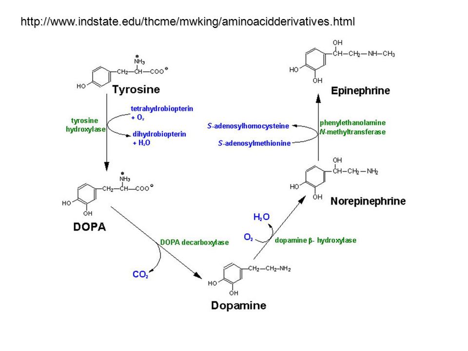
Synthesis of Neurotransmitters

34
http://www.indstate.edu/thcme/mwking/aminoacidderivatives.html
35
Pathway for serotonin and melatonin synthesis from tryptophan. Abbreviations: THP = tryptophan hydroxylase, DHPR = dihydropteridine reductase, H2B = dihydrobiopterin, H4B = tetrahyrobiopterin, 5-HT = 5-hydroxytryptophan, AADC = aromatic L-amino acid decarboxylase, SNA = serotonin N-acetylase, HOMT = hydroxyindole-O-methyltransferase.

37
Transmitter release SNARE: soluble NSF attachment receptor NSF NSF: N- ethylmaleimide sensitive fusion protein SNARE proteins on two joining membranes (usually a vesicle and a target membrane such as the plasma membrane) form a tight complex. The role of NSF is to undo these SNARE complexes once membrane fusion has occurred. The dissociated SNAREs can then be recycled for reuse in further rounds of membrane fusion. SNAP-25 (synaptosome- associated protein of 25,000 daltons) Soluble NSF attachment protein (-SNAP) are thought to be soluble factors that transiently bind and disassemble SNAP receptor complex during exocytosis in neuronal and endocrine cells.
 Soluble NSF attachment protein (-SNAP) are thought to be soluble factors that transiently bind and disassemble SNAP receptor complex during exocytosis in neuronal and endocrine cells..")
38
http://www.neuro.wustl.e du/neuromuscular/pathol /snare.htm Synaptobrevin: v-SNARE Synaptotagmin: Calcium sensor Syntaxin: t-SNARE SNAP receptors implicated in vesicle targeting and fusion. Sollner T, Whiteheart SW, Brunner M, Erdjument-Bromage H, Geromanos S, Tempst P, Rothman JE. Sollner TWhiteheart SWBrunner MErdjument-Bromage H Geromanos STempst PRothman JE The N-ethylmaleimide-sensitive fusion protein (NSF) and the soluble NSF attachment proteins (SNAPs) appear to be essential components of the intracellular membrane fusion apparatus. An affinity purification procedure based on the natural binding of these proteins to their targets was used to isolate SNAP receptors (SNAREs) from bovine brain. Remarkably, the four principal proteins isolated were all proteins associated with the synapse, with one type located in the synaptic vesicle and another in the plasma membrane, suggesting a simple mechanism for vesicle docking. The existence of numerous SNARE- related proteins, each apparently specific for a single kind of vesicle or target membrane, indicates that NSF and SNAPs may be universal components of a vesicle fusion apparatus common to both constitutive and regulated fusion (including neurotransmitter release), in which the SNAREs may help to ensure vesicle-to-target specificity.
 and the soluble NSF attachment proteins (SNAPs) appear to be essential components of the intracellular membrane fusion apparatus. An affinity purification procedure based on the natural binding of these proteins to their targets was used to isolate SNAP receptors (SNAREs) from bovine brain. Remarkably, the four principal proteins isolated were all proteins associated with the synapse, with one type located in the synaptic vesicle and another in the plasma membrane, suggesting a simple mechanism for vesicle docking. The existence of numerous SNARE- related proteins, each apparently specific for a single kind of vesicle or target membrane, indicates that NSF and SNAPs may be universal components of a vesicle fusion apparatus common to both constitutive and regulated fusion (including neurotransmitter release), in which the SNAREs may help to ensure vesicle-to-target specificity..")
39
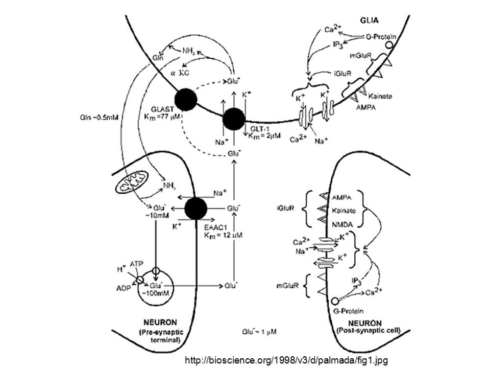
http://bioscience.org/1998/v3/d/palmada/fig1.jpg
40
http://bioscience.org/1998/v3/d/palmada/d701-718.htm

Hasonló előadás


 Kürti Jenő Koltai János (helyettesítés) ELTE Biológiai.>")


