Előadást letölteni
Az előadás letöltése folymat van. Kérjük, várjon

1
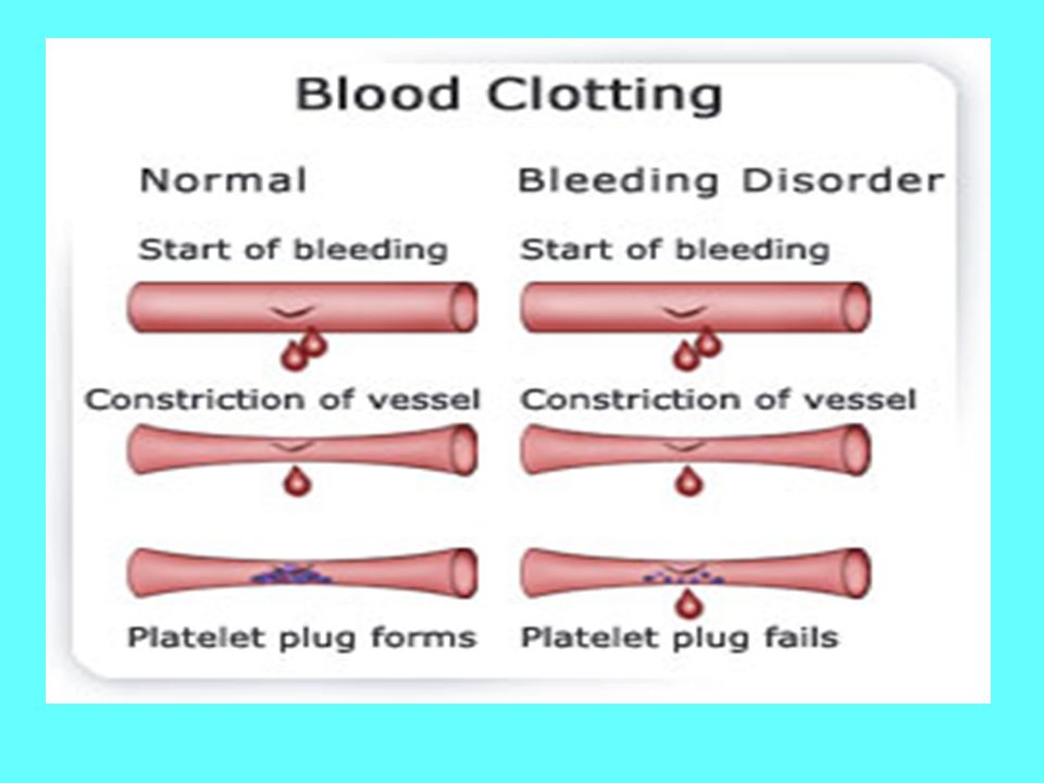
Hemosztázis (Vérzéscsillapítás)
A hemosztázis feladata : A vér folyékony állapotban tartása az érpályán belül, érfali sérülés esetén az elvérzés megakadályozása, majd az érfali integritás helyreállítása. Ennek fázisai: 1. Érösszehúzódás (vaszkuláris fázis) (kb. 30 sec) 2 Vérlemezke fázis (trombocita dugó képződés) (kb. 3-7 perc) 3. Plazma fázis (fibrinképződés) (kb perc) 4. Fibrinolízis (48-72 óra)
 (kb. 30 sec) 2 Vérlemezke fázis (trombocita dugó képződés) (kb. 3-7 perc) 3. Plazma fázis (fibrinképződés) (kb perc) 4. Fibrinolízis (48-72 óra)")
2
Vérzékenység Fokozott alvadási készség

3
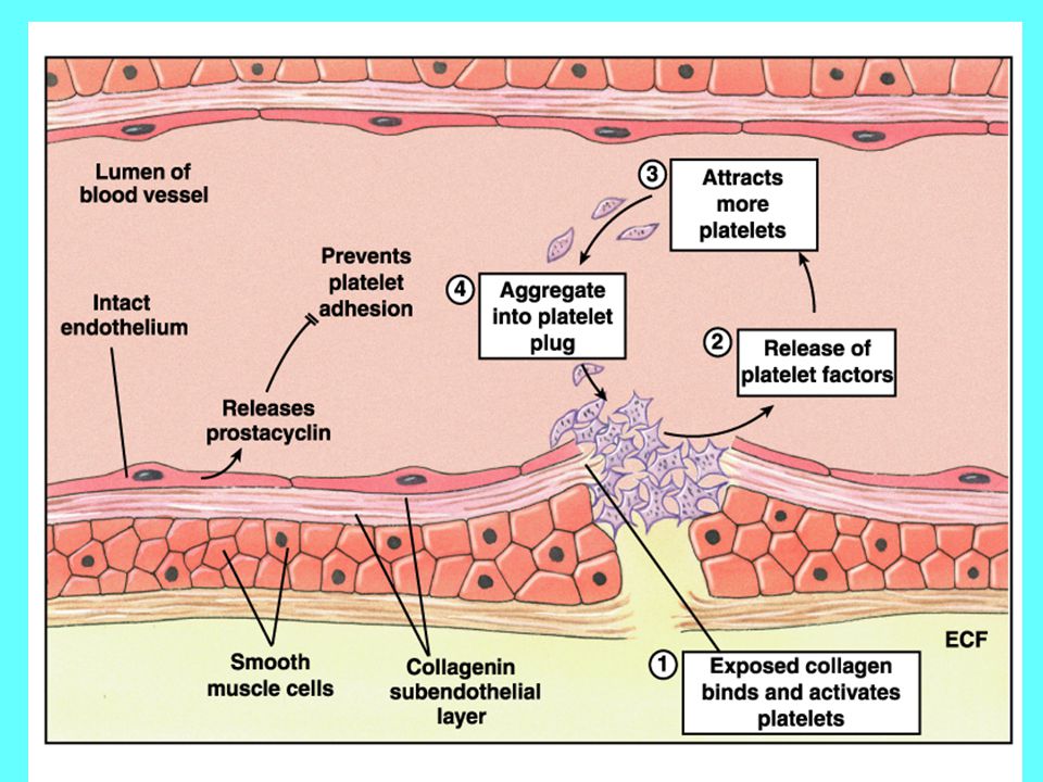
Ér (vaszkuláris) - fázis
 - fázis")
5
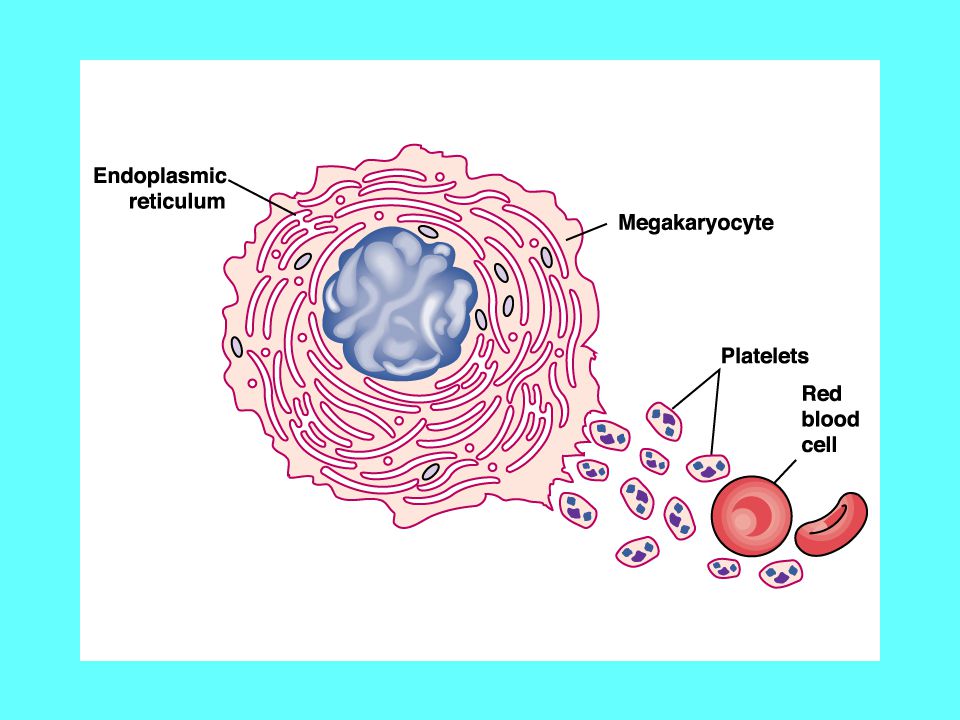
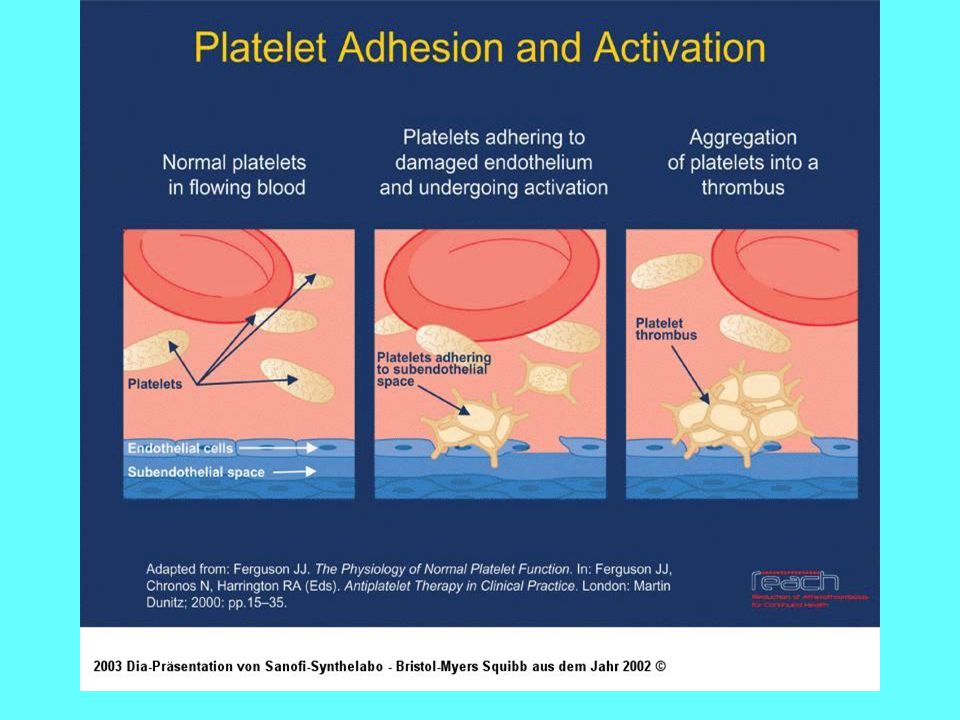
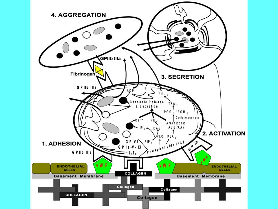
Vérlemezke (trombocita) - fázis
 - fázis")
10
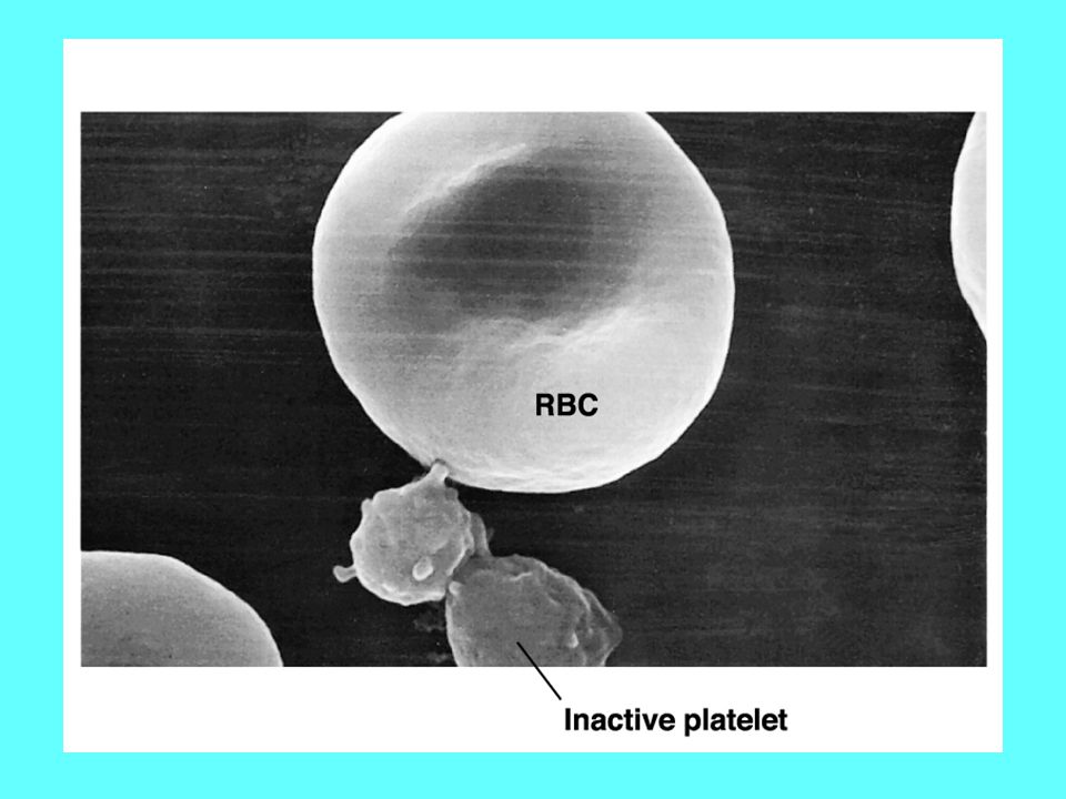
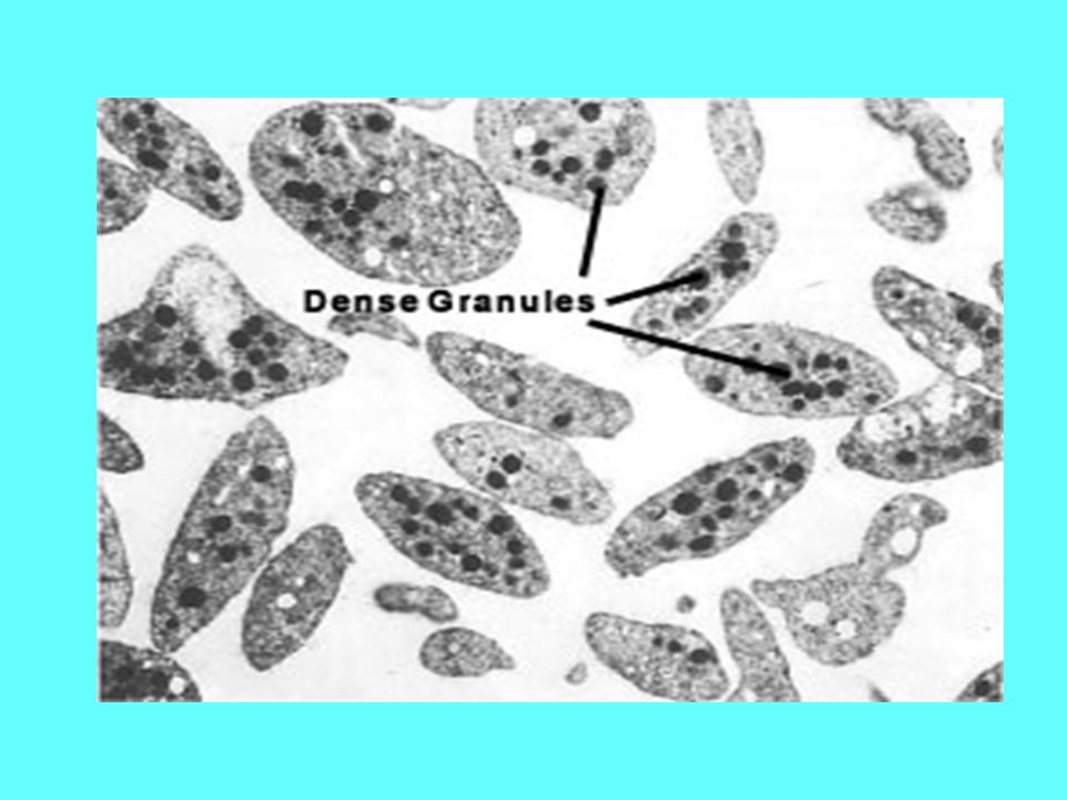
Nyugvó vérlemezkék

11
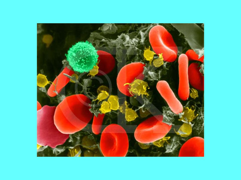
Aktiválódott vérlemezkék

17
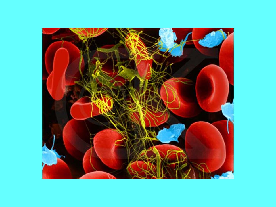
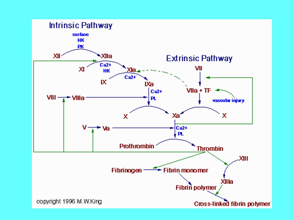
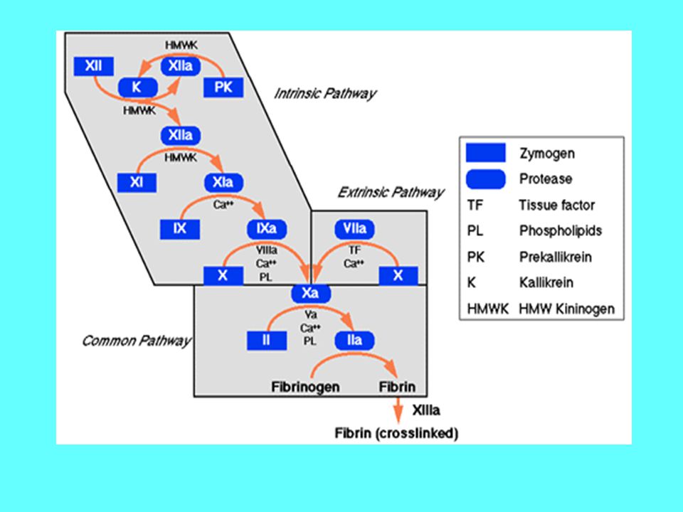
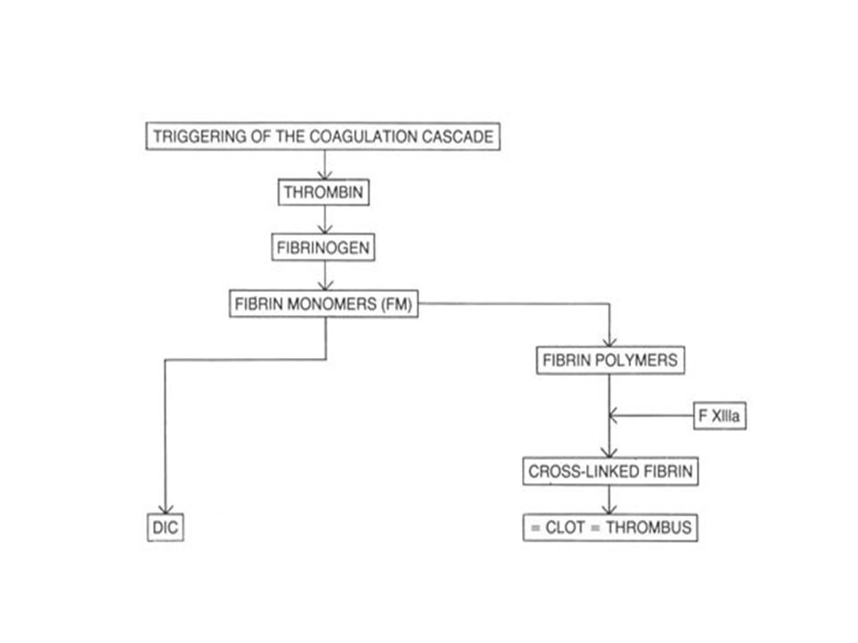
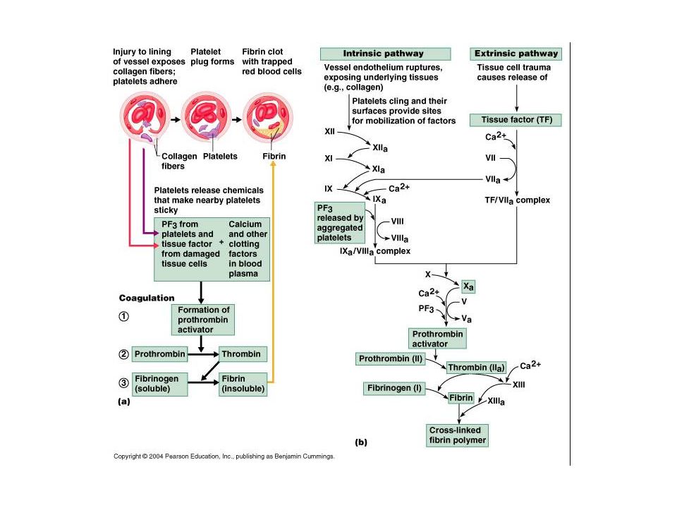
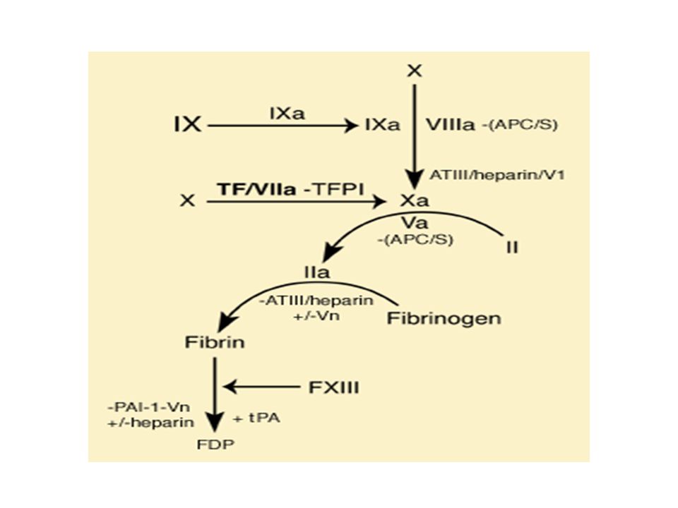
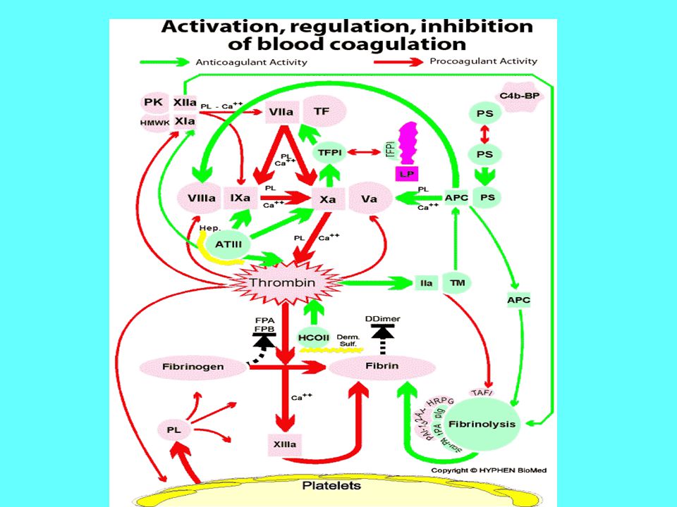
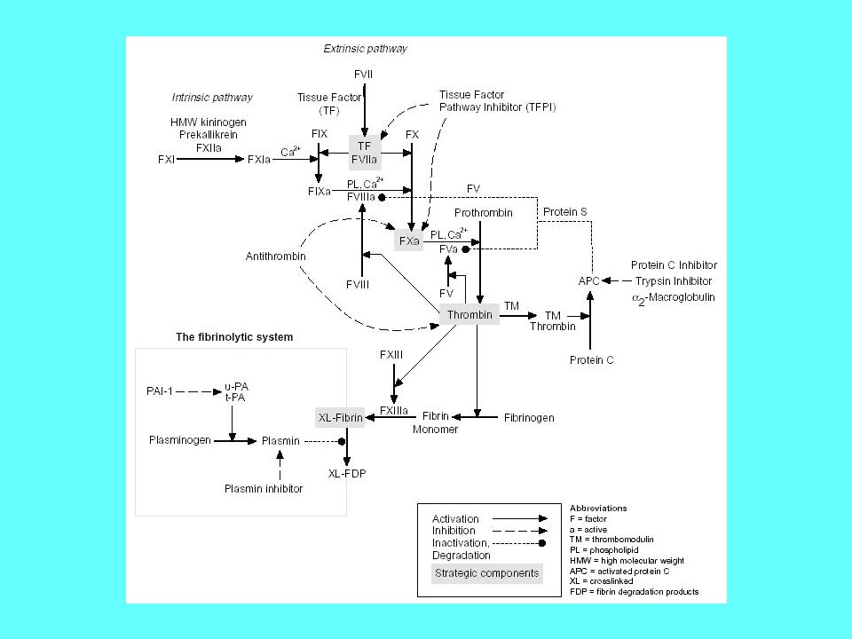
Plazma - fázis (koaguláció)
")
20
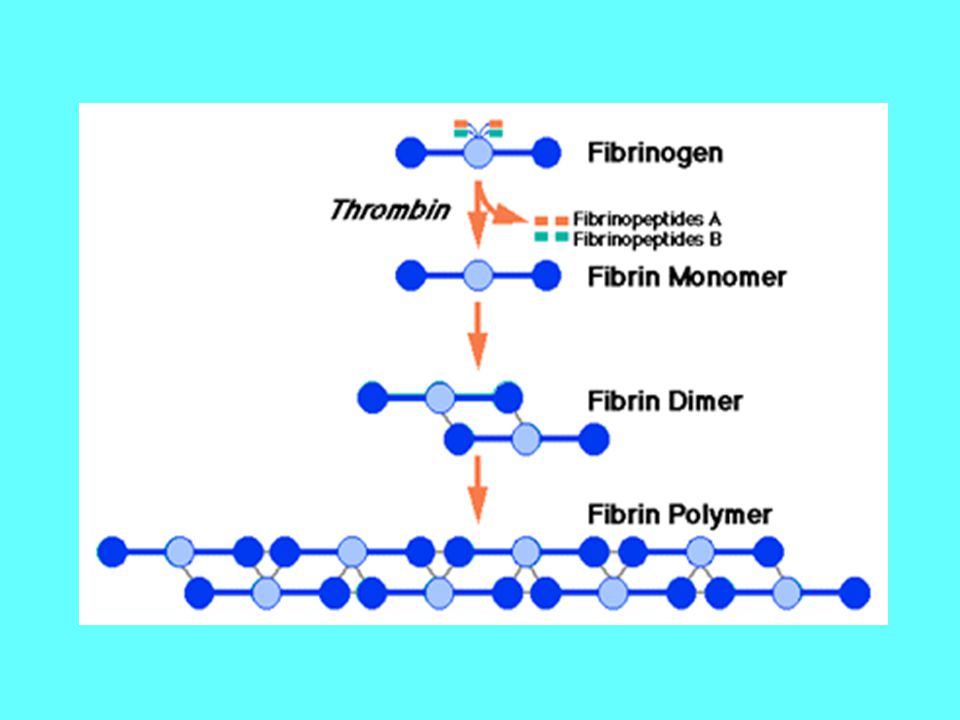
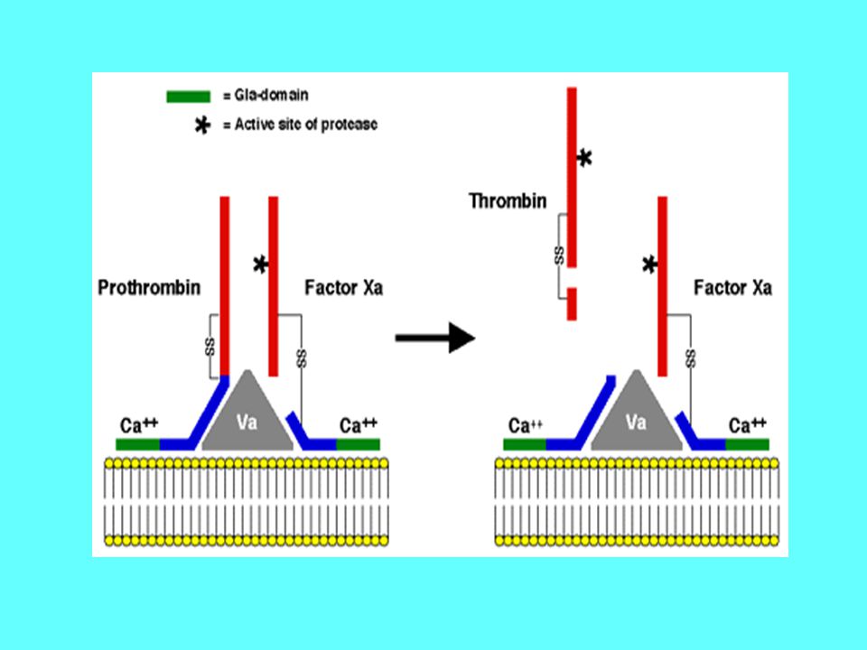
fibrinogen and thrombin
The heart of the reaction involves just two molecules: fibrinogen and thrombin Thrombin Fibrinogen Fibrin Thrombin removes A’s and B’s, converting fibrinogen to fibrin. Fibrin proteins clump together due to the affinity of a and b for the sticky crevices left by A’s and B’s excision.

24
Rube Goldberg machines
Pencil Sharpener Open window (A) and fly kite (B). String (C) lifts small door (D) allowing moths (E) to escape and eat red flannel shirt (F). As weight of shirt becomes less, shoe (G) steps on switch (H) which heats electric iron (I) and burns hole in pants (J). Smoke (K) enters hole in tree (L), smoking out opossum (M) which jumps into basket (N), pulling rope (O) and lifting cage (P), allowing woodpecker (Q) to chew wood from pencil (R), exposing lead. Emergency knife (S) is always handy in case opossum or the woodpecker gets sick and can't work.
 and fly kite (B). String (C) lifts small door (D) allowing moths (E) to escape and eat red flannel shirt (F). As weight of shirt becomes less, shoe (G) steps on switch (H) which heats electric iron (I) and burns hole in pants (J). Smoke (K) enters hole in tree (L), smoking out opossum (M) which jumps into basket (N), pulling rope (O) and lifting cage (P), allowing woodpecker (Q) to chew wood from pencil (R), exposing lead. Emergency knife (S) is always handy in case opossum or the woodpecker gets sick and can t work.")
25
Irreducible Complexity
The modern form of The Argument from Personal Incredulity: Irreducible Complexity "An irreducibly complex system cannot be produced directly by numerous, successive, slight modifications of a precursor system, because any precursor to an irreducibly complex system that is missing a part is by definition nonfunctional Since natural selection can only choose systems that are already working, then if a biological system cannot be produced gradually it would have to arise as an integrated unit, in one fell swoop, for natural selection to have anything to act on."

26
Invertebrates DO have fibrinogen.
Parastichopus parvimensis (warty sea cucumber)
")
27
kb. 450 millió évvel ezelőtt.
Orsóhal (ingola) az első gerinces állat, amelyben megjelent a fibrinogén kb. 450 millió évvel ezelőtt.
 az első gerinces állat, amelyben megjelent a fibrinogén. kb. 450 millió évvel ezelőtt.")
28
Based upon the sequence comparisons, a scenario for when clotting proteins made their appearance
Doolittle, R. F., and Feng, D. F., (1987) Cold Spring Harbor Symposia on Quantitative Biology 52:
 Cold Spring Harbor Symposia on Quantitative Biology 52:")
29
A model for the evolution of blood clotting
Most serine proteases, including trypsin and thrombin, are auto-catalytic. The inactive form of the protease (A) is changed into the active form (A*) when two things happen: it is bound to tissue factor (TF) and it is activated by tissue proteases, including our protease itself (that's the autocatalytic part). This means - and this is important - that our protease is actually involved in cutting two things: Fibrinogen, and also itself, converting A's inactive precursor protein into A*. Ken Miller,
 is changed into the active form (A*) when two things happen: it is bound to tissue factor (TF) and it is activated by tissue proteases, including our protease itself (that s the autocatalytic part). This means - and this is important - that our protease is actually involved in cutting two things: Fibrinogen, and also itself, converting A s inactive precursor protein into A*. Ken Miller,")
30
A gene duplication occurs in the gene for our protease, producing a new (B) version of the gene:
Proteins A and B are identical. Each can bind to TF, each can cleave fibrinogen into fibrin, and each can activate itself or its sister serum protease. So nothing has really changed - we've just got two copies of the same gene. Ken Miller,

31
Network rearrangements
A mutation in the active site of B changes its behavior, making it a little less likely to cut fibrinogen and a little more likely to activate protease A. But why would natural selection favor a mutation like this in B's active site? Ken Miller,

32
The evolution of the meaning of protein function
traditional view post-genomic view Eisenberg et al. Nature : 823-6

33
Network expansion by gene duplication
b shows a small protein interaction network (blue) and the genes that encode the proteins (green). When cells divide, occasionally one or several genes are copied twice into the offspring’s genome (illustrated by the green and red circles). This induces growth in the protein interaction network because now we have an extra gene that encodes a new protein (red circle). The new protein has the same structure as the old one, so they both interact with the same proteins. Ultimately, the proteins that interacted with the original duplicated protein will each gain a new interaction to the new protein. Therefore proteins with a large number of interactions tend to gain links more often, as it is more likely that they interact with the protein that has been duplicated. This is a mechanism that generates preferential attachment in cellular networks. Indeed, in the example that is shown it does not matter which gene is duplicated, the most connected central protein (hub) gains one interaction. In contrast, the square, which has only one link, gains a new link only if the hub is duplicated. Barabasi & Oltvai. NRG. (2004)
 and the genes that encode the proteins (green). When. cells divide, occasionally one or several genes are copied twice into the offspring’s genome (illustrated by the green and red circles). This induces growth in the protein interaction network because now we have an extra gene that encodes a new protein (red circle). The new protein has the same structure as the old one, so they both interact with the same proteins. Ultimately, the proteins that interacted with the original duplicated protein will each gain a new interaction to the new protein. Therefore proteins with a large number of interactions tend to gain links more often, as it is more likely that they interact with the protein that has been duplicated. This is a mechanism that generates preferential attachment in cellular networks. Indeed, in the example that is shown it does not matter which gene is duplicated, the most connected central protein (hub) gains one interaction. In contrast, the square, which has only one link, gains a new link only if the hub is duplicated. Barabasi & Oltvai. NRG. (2004)")
34
Hálózatok evolúciója Gén duplikáció Pontmutáció (megváltozott funkció) 3. Új szabályozó mechanizmusok érvényre jutása

35
Yeast protein-protein interaction networks
the phenotypic effect of removing the corresponding protein: Lethal Slow-growth Non-lethal Unknown Jeong et al. Nature 411, (2001)
")
36
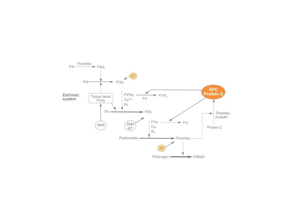
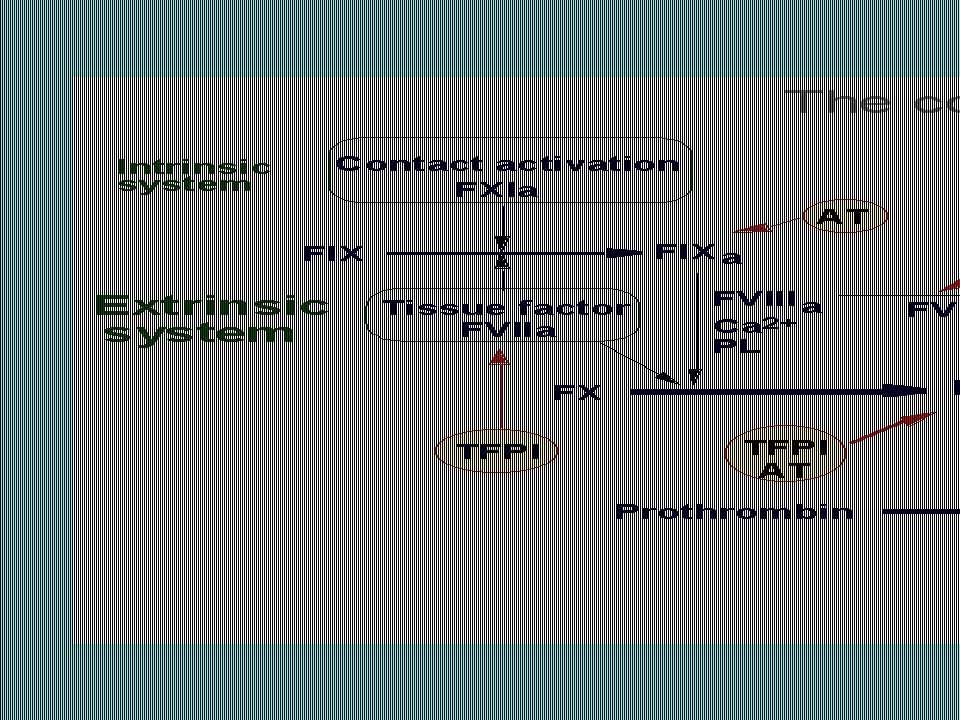
A véralvadási rendszer inhibitorai
Antitrombin Heparin kofaktor II α1-antitripszin α2-makroglobulin Tissue factor pathway inhibitor (TFPI) Trombomodulin Protein C rendszer Protein S Heparin Dermatán szulfát
 Trombomodulin. Protein C rendszer. Protein S. Heparin. Dermatán szulfát.")
37
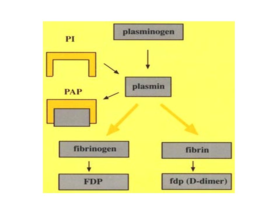
Fibrinolízis

38
degradation products (FDP)
Fibrinolytic Pathway PAI-1 Plasminogen Tissue Plasminogen Activator (t-PA) Urokinase (uPA) Exogenous: streptokinase Plasmin Inhibitor XL-Fibrin, fibrinogen Plasmin XL- fibrin degradation products (FDP)
 Urokinase (uPA) Exogenous: streptokinase. Plasmin Inhibitor. XL-Fibrin, fibrinogen. Plasmin. XL- fibrin. degradation products (FDP)")
39
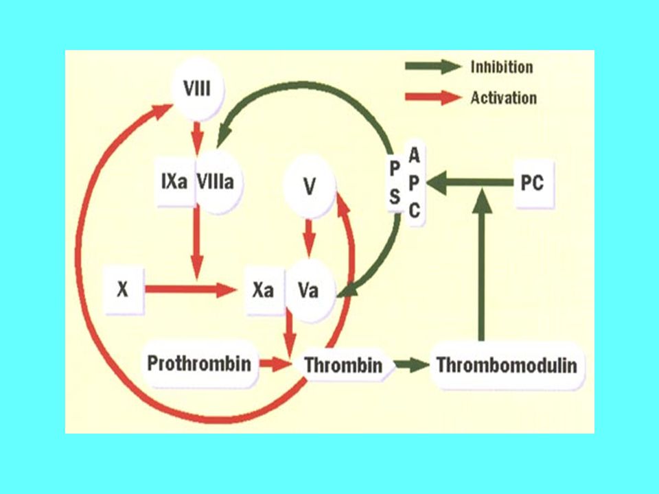
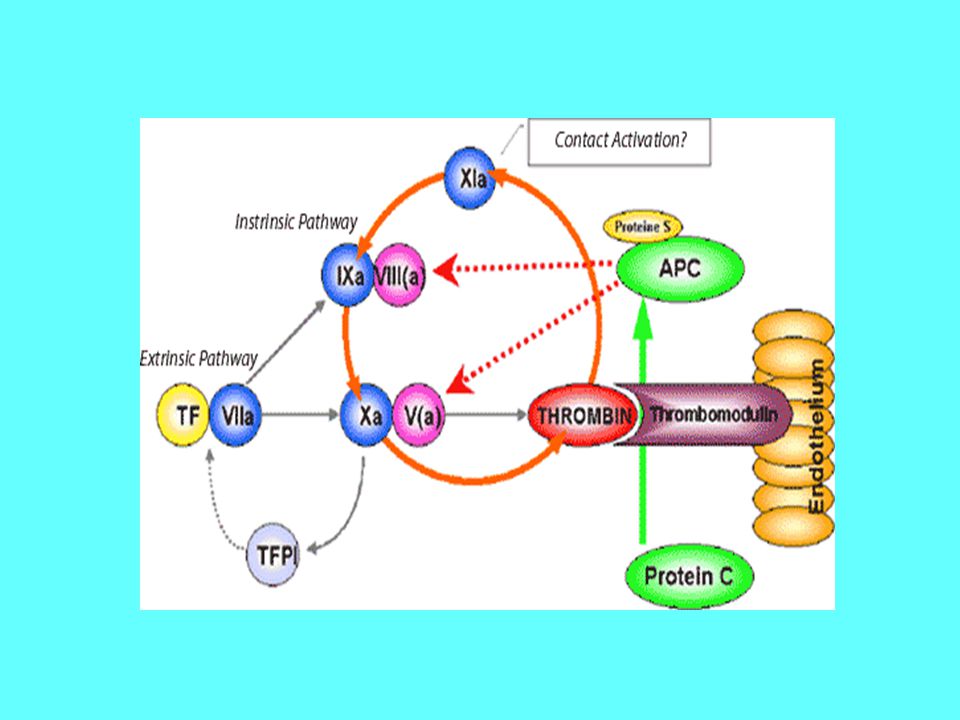
Anticoagulation Pathways – Protein C
Protein C Inhibitor (PAI-3) Trypsin Inhibitor a2-Macroglobulin FX Prothrombin APC Protein S FVIIIa FV PL, Ca2+ FXa FVa Protein C Thrombin Thrombo-modulin Fibrinogen Fibrin
 Trypsin Inhibitor. a2-Macroglobulin. FX. Prothrombin. APC. Protein S. FVIIIa. FV. PL, Ca2+ FXa. FVa. Protein C. Thrombin. Thrombo-modulin. Fibrinogen Fibrin.")
41
Anticoagulation Pathways - Antithrombin
FX TF FVIIa Prothrombin TFPI PL FXa Heparin (cofactor) Va Antithrombin III Thrombin
 Va. Antithrombin III. Thrombin.")
42
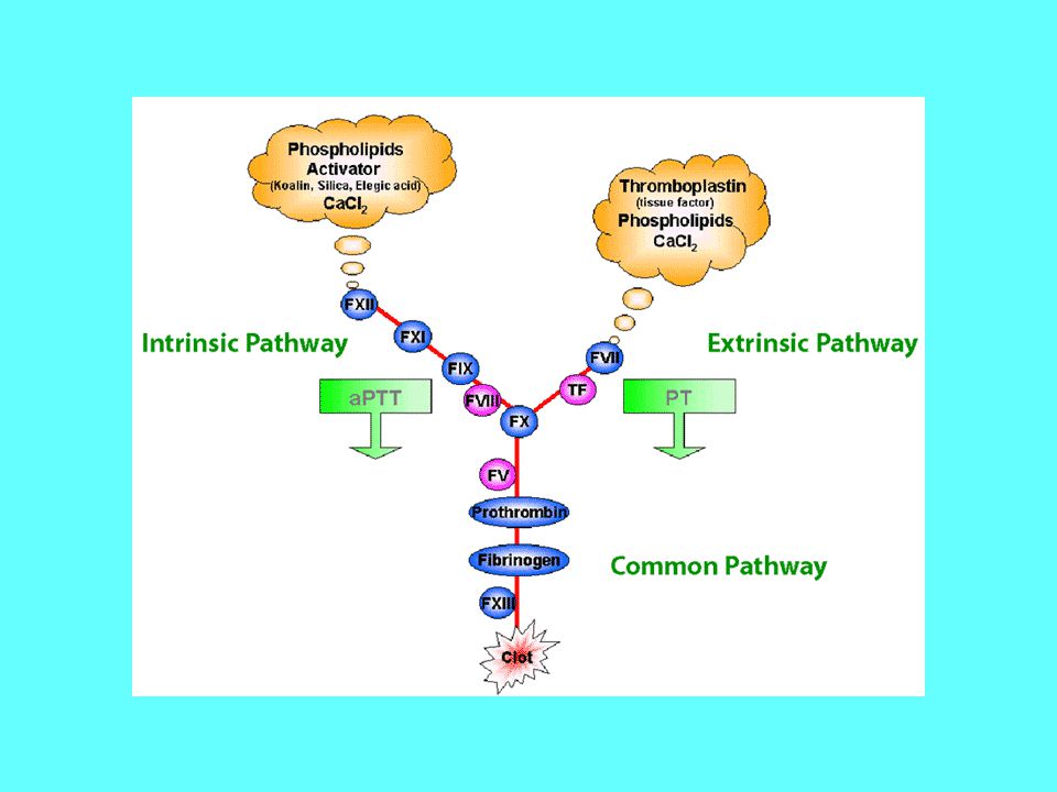
Véralvadási szűrő tesztek
Vérzési idő Protrombin idő (PI) (extrinszik út faktorai) Aktivált parciális tromboplasztin idő (APTI) (intrinszik út faktorai) Trombin idő (TI) Fibrinogén koncentráció D-dimer teszt
 (extrinszik út faktorai) Aktivált parciális tromboplasztin idő (APTI) (intrinszik út faktorai) Trombin idő (TI) Fibrinogén koncentráció. D-dimer teszt.")
43
Vérzékenységi betegségek
Vérlemezkékkel kapcsolatos zavarok: Csökkent szám (trombocitopéniák) „ funkció (trombocitopátiák) Alvadási zavarok (koagulopátiák) Komplex zavarok Egyéb
 „ funkció (trombocitopátiák) Alvadási zavarok (koagulopátiák) Komplex zavarok. Egyéb.")
44
Hemofiliák Hemofilia A (VIII Faktor képzési zavara, X-kromoszóma)
Hemofilia B (IX Faktor képzési zavara, X-kromoszóma) Hemofilia C (XI Faktor képzési zavara, 4. kromoszóma) Hemofilia A legjellemzőbb tünetei: (gyakoriság: 20/100 ezer fiú) Ismétlődő izületi és izomközi bevérzések, bőralatti vérzések, műtétek és traumák utáni szűnni nem akaró vérzések. Izületi deformitások Súlyos: VIIIF szint < 1%, mérsékelt: 1-4%, enyhe: 5-25% A hemofilia B tünetei hasonlóak a Hemofilia A-hoz A hemofilia C tünetei enyhébbek a másik kettőnél
 Hemofilia C (XI Faktor képzési zavara, 4. kromoszóma) Hemofilia A legjellemzőbb tünetei: (gyakoriság: 20/100 ezer fiú) Ismétlődő izületi és izomközi bevérzések, bőralatti vérzések, műtétek és traumák utáni szűnni nem akaró vérzések. Izületi deformitások. Súlyos: VIIIF szint < 1%, mérsékelt: 1-4%, enyhe: 5-25% A hemofilia B tünetei hasonlóak a Hemofilia A-hoz. A hemofilia C tünetei enyhébbek a másik kettőnél.")
46
von Willebrand betegség
Három formája ismeretes: A vWF működése normális, de a mennyisége kevés A vWF mennyisége normális, de a működése kóros A vWF csaknem teljesen hiányzik Tünetei: bőr és nyálkahártya vérzések (enyhe traumák után), orrvérzés, foghúzás utáni elhúzódó vérzések. Típustól függően enyhétől az igen súlyosig terjedhetnek.
, orrvérzés, foghúzás utáni elhúzódó vérzések. Típustól függően enyhétől az igen súlyosig terjedhetnek.")
47
A trombózis veleszületett hajlamosító tényezői
A protein C képzés zavarai Az FV Leiden mutáció (Arg506Gln) Emelkedett FVIII szint (>150%) Emelkedett FXI szint A fibrinogén képződés mennyiségi és minőségi zavarai A fibrinolítikus enzim-rendszer működésének zavarai
 Emelkedett FVIII szint (>150%) Emelkedett FXI szint. A fibrinogén képződés mennyiségi és minőségi zavarai. A fibrinolítikus enzim-rendszer működésének zavarai.")
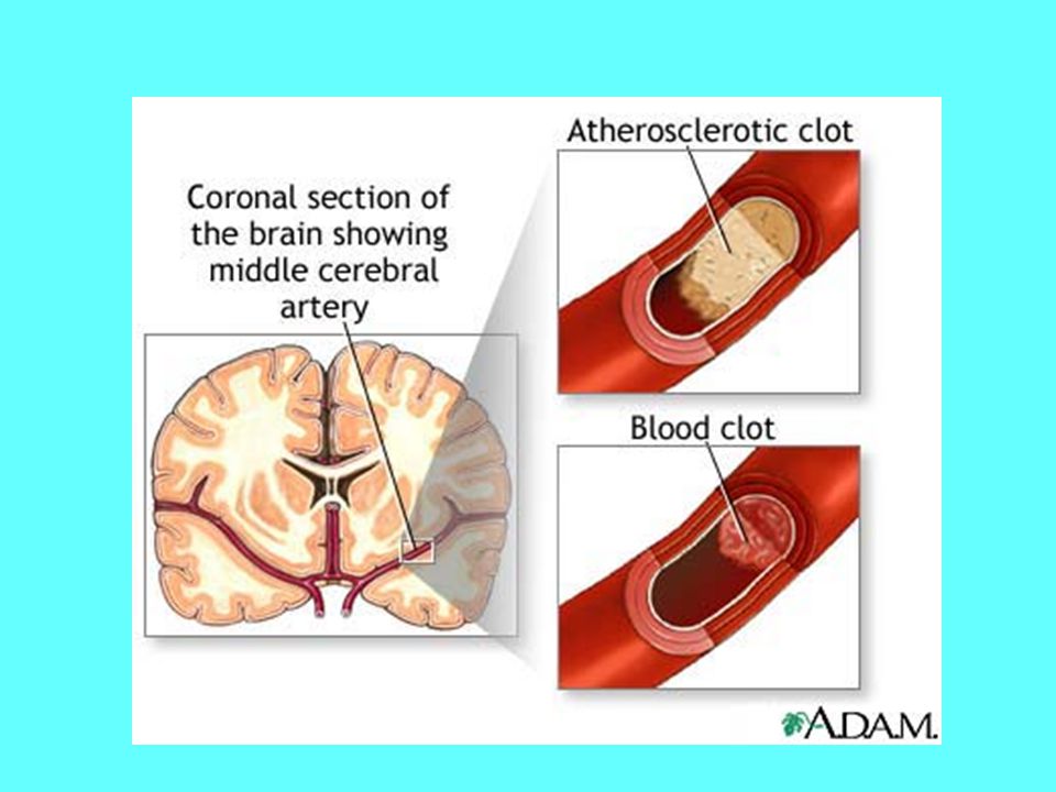
48
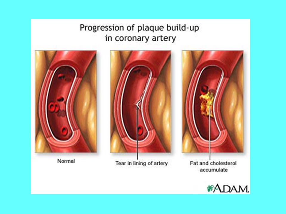
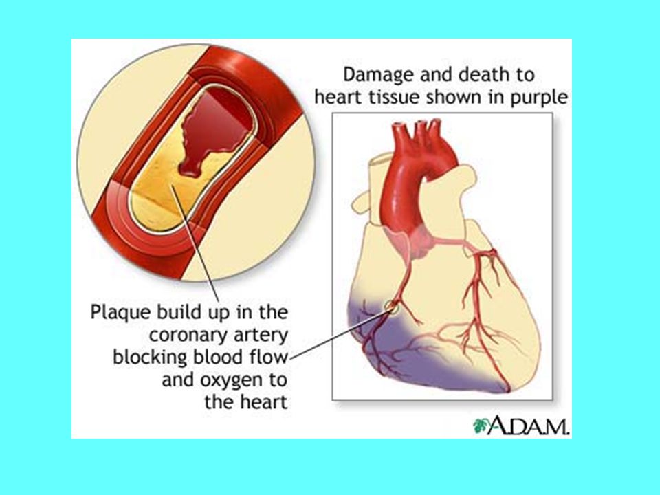
A trombózis szerzett hajlamosító tényezői
Érelmeszesedés (az artériás oldalt érinti) Rosszindulatú betegségek (főleg a vénás oldalt érintik) Hemolízis Fokozott viszkozitás Általános sebészeti műtétek Ortopédiai műtétek Általános sebészeti műtétek Terhesség Orális fogamzásgátlók, hormonpótló kezelés Tartós immobilitás Elhízás Antifoszfolipd szindroma
 Rosszindulatú betegségek (főleg a vénás oldalt érintik) Hemolízis. Fokozott viszkozitás. Általános sebészeti műtétek. Ortopédiai műtétek. Általános sebészeti műtétek. Terhesség. Orális fogamzásgátlók, hormonpótló kezelés. Tartós immobilitás. Elhízás. Antifoszfolipd szindroma.")
51
„Don’t worry about your heart, it will last you
as long you live” (W.C. Fields)
")
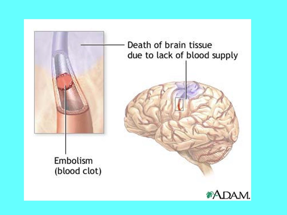
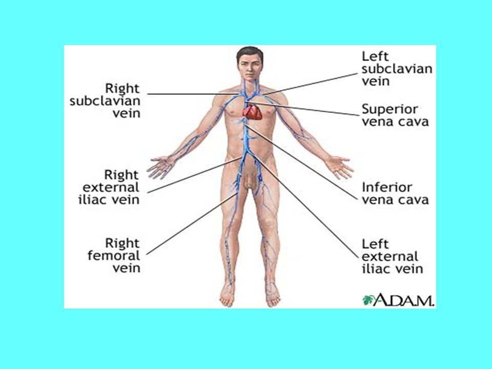
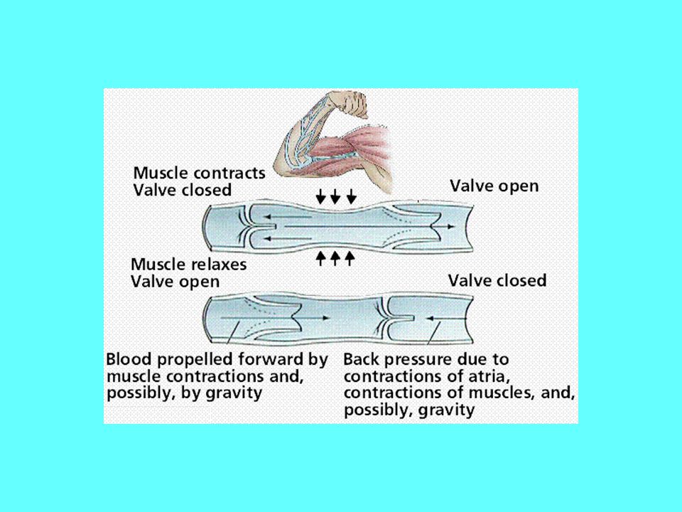
57
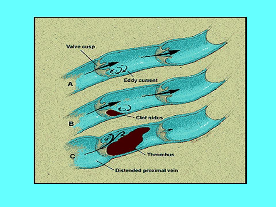
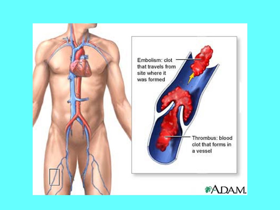
Mély vénás trombózis

59
Everybody talks about it, nobody understands it. JH Levy 2000
LOVE = HEMOSTASIS Everybody talks about it, nobody understands it. JH Levy 2000 In summary, hemostasis and hemostatic mechanisms are complex physiologic responses that are important for clinicians to understand when managing critical ill patients.

68
Anticoagulation proteins: Protein C, Protein S, Antithrombin III
Coagulation Cascade Intrinsic Pathway Extrinsic Pathway Contact Activation IX TF Pathway Tissue Factor + VII X XI TF-VIIa Prekallikrein HMW Kininogen Ca2+ PL Common Pathway XIIa Prothrombin XIa PL, Ca2+ (Tenase) IXa VIIIa PL, Ca2+ Xa XIII Va Anticoagulation proteins: Protein C, Protein S, Antithrombin III (Prothrombinase) Thrombin XIIIa Fibrinogen Fibrin Monomer Fibrin Polymer
 IXa. VIIIa. PL, Ca2+ Xa. XIII. Va. Anticoagulation proteins: Protein C, Protein S, Antithrombin III. (Prothrombinase) Thrombin. XIIIa. Fibrinogen. Fibrin. Monomer. Fibrin. Polymer.")
79
Most of the enzymes involved in clotting are serine proteases
The serine proteases are homologous. They are also homologous to the pancreatic serine proteases trypsin, chymotrypsin, and elastase. The N-terminal segments are thought to be responsible, at least in part, for the specificities of the proteolytic blood clotting factors. The Evolution of Vertebrate Blood Clotting By Kenneth Miller,

80
The blood clotting system looks like a Rube Goldberg machine
Is it Irreducibly Complexity? From Stryers’ Biochemistry

82
The domain architecture of blood coagulation proteins reveals a history of exon shuffling
Peer Bork’s Modules page:

83
Fibrinogen is composed of two each of three homologous polypeptide chains (a, b, g)
")
84
Szöveti faktor + VII Faktor komplex

85
B A A+ B+ TF TF Fibrinogén Fibrin B B+ TF A A+ Fibrinogén Fibrin

Hasonló előadás


 MORE?” symposium Washington.>")


