Előadást letölteni
Az előadás letöltése folymat van. Kérjük, várjon

1
Anaemia gyermekkorban Molnár Dénes Gyermekklinika

2
Cél Anaemia okainak áttekintése Anaemia diagnózisának felállításának megközelítése Vashiányos anaemia diagnosisa és kezelése Rövid tárgyalása a gyermekkori anaemiák egyéb okainak

3
A vörösvértest Élettartam: 120 days – 60-90 days in term & 35-50 days in preterm babies Termelődés –Regulated by epo (other hemopoietic factors. Colony- stimulating factors, interleukins, thrombopoietin are not mentioned here) Produced by kidneys in response to low O2 –Epo stimulates marrow to make RBC precursors –Needs iron, B12, folate, and amino acid Destrukció –When old or damaged, taken up by spleen
 Produced by kidneys in response to low O2 –Epo stimulates marrow to make RBC precursors –Needs iron, B12, folate, and amino acid Destrukció –When old or damaged, taken up by spleen.")
4
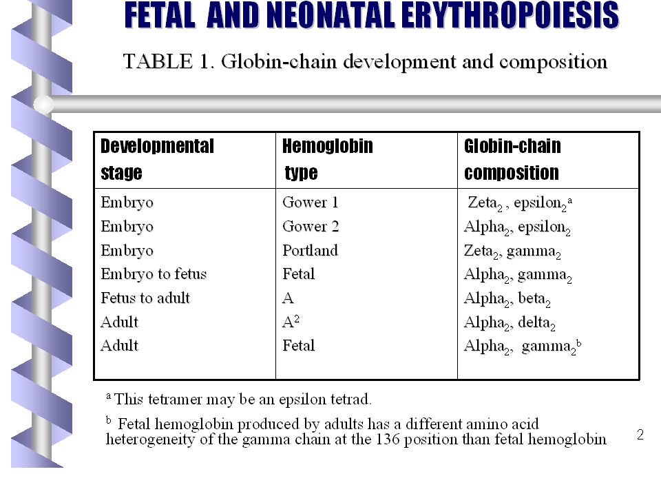
A vörösvértest Membrán Enzimek Hemoglobin –Heme (4 heme groups per Hb molecule) Mediates binding of O2 by Hb –Globin Protein that surrounds and protects heme molecule 2 alpha and 2 beta genes (adult Hb)
 Mediates binding of O2 by Hb –Globin Protein that surrounds and protects heme molecule 2 alpha and 2 beta genes (adult Hb)")
6
Fontosabb terminológia Hb –Vvt hb koncentrációja a vérben Ht (%) –A vér alakos elemeinek frakcionált volumene a vérben MCV –A vvt átlagos térfogata Microcyter, macrocyter, normocyter MCHC –Kalkulált érték (Hb/Ht) Alacsony MCHC hypochromiára utal
 –A vér alakos elemeinek frakcionált volumene a vérben MCV –A vvt átlagos térfogata Microcyter, macrocyter, normocyter MCHC –Kalkulált érték (Hb/Ht) Alacsony MCHC hypochromiára utal")
7
Fontosabb terminológia RDW ( Red Cell Distribution Width) –Vvt nagyságának variabilitását mutatja; normál értéke 11-15 Ferritin –Vas raktározott formája 1 ng ferritin: 10 mg raktározott vas Transferrin –Vas transzportját végzi Reticulociták –Fiatal vvt
 –Vvt nagyságának variabilitását mutatja; normál értéke Ferritin –Vas raktározott formája 1 ng ferritin: 10 mg raktározott vas Transferrin –Vas transzportját végzi Reticulociták –Fiatal vvt")
8
Anaemia okai Csökkent produkció Fokozott destrukció Vérvesztés

9
Csökkent vvt termelés Defective heme synthesis –Iron deficiency, anemia of chronic disease, lead poisoning Defective globin synthesis –Alpha and beta thalassemia Defective DNA synthesis –Nutrient deficiencies (B12, folate) Impaired epo production –Renal disease
 Impaired epo production –Renal disease")
10
Csökkent vvt termelés Marrow failure –Aplastic anemia congenital Fanconi anemia is an autosomal recessive disorder affecting all bone marrow elements and associated with cardiac, renal, and limb malformations as well as dermal pigmentary changes. (congenital) acquired –Red cell aplasia Congenital (Diamond-Blackfan) Acquired (Transient erythroblastopenia of childhood) Marrow replacement –Malignancy, myelofibrosis
 acquired –Red cell aplasia Congenital (Diamond-Blackfan) Acquired (Transient erythroblastopenia of childhood) Marrow replacement –Malignancy, myelofibrosis.")
11
Fokozódó destrukció Extracellular –Antibody mediated –Microangiopathic; HUS, DIC –Drugs, toxins –Hypersplenism

12
Fokozott pusztulás Intracelluláris –RBC membrán defektusok HS (hereditary spherocytosis, stomatocytosis [For as yet unknown reasons, the cells take on an abnormal shape, resembling a mouth or 'stoma‘]), HE (hereditary eliptocytosis) –Enzim defektusok PK (pyruvate kinase), G6PD –Hemglobinopathiák Sickle cell, thalassemia
![Fokozott pusztulás Intracelluláris –RBC membrán defektusok HS (hereditary spherocytosis, stomatocytosis [For as yet unknown reasons, the cells take on an abnormal shape, resembling a mouth or stoma‘]), HE (hereditary eliptocytosis) –Enzim defektusok PK (pyruvate kinase), G6PD –Hemglobinopathiák Sickle cell, thalassemia](http://images.slideplayer.hu/34/10194957/slides/slide_12.jpg "Fokozott pusztulás Intracelluláris –RBC membrán defektusok HS (hereditary spherocytosis, stomatocytosis [For as yet unknown reasons, the cells take on an abnormal shape, resembling a mouth or stoma‘]), HE (hereditary eliptocytosis) –Enzim defektusok PK (pyruvate kinase), G6PD –Hemglobinopathiák Sickle cell, thalassemia")
13
Vérvesztés Akut vérzés Krónikus vérvesztés

14
Anaemia Definíció –Hb concentráció > 2 SD a normál populációs átlag alatt Normál értékek korral változnak

15
Életkori változások ÉletkorÚjszülött2 hetes3 hónapos6hó – 6 év7-12 év Hb (g/l)168 (137-210) 165 (130-200) 120 (95-145) 120 (105-140) 130 (110-160) Hct (%)55 (45-65)50 (42-66)36 (31-41)37 (33-42)38 (34-40) MCV (fl) lowest 11070-7476-80 Reticuloc ytes (%) 51111
168 ( ) 165 ( ) 120 (95-145) 120 ( ) 130 ( ) Hct (%)55 (45-65)50 (42-66)36 (31-41)37 (33-42)38 (34-40) MCV (fl) lowest Reticuloc ytes (%) 51111")
16
Physiologiás anaemia Hb csökkenés 3 hónapos kor környékén Háttérben zajló fiziológiás változások –At birth, O2 sat rises sharply 65% in utero, nearly 100% when comes out –Decrease in epo production –Fetal RBC have shorter survival time (60 days)
")
17
Anaemia diagnózisa Anamnézis Fizikális vizsgálat Labor és egyéb vizsgálatok

18
Anamnézis Anyai anamnézis –Anaemia terhesség alatt –Terhesség és szülés Családi anamnézis –Etnikai hovatartozás –Anaemia –Sárgaság, epekőbetegség, cholecystectomia –Transfusio –Splenomegalia

19
Anamnézis Páciens –Sárgaság születés utáni időszakban –Diétás szokások –Gyógyszerszedés –Infekciók a közelmúltban –Krónikus betegségek –Bőrvérzések, vérzések –Pica

20
Fizikális vizsgálat General appearance –Pallor, jaundice, bruising Head and neck –Pale mucous membranes and conjunctiva –Angular stomatitis –Adenopathy Cardiac –Tachycardia, heart failure if severe –Heart murmur Abdomen –Organomegaly

21
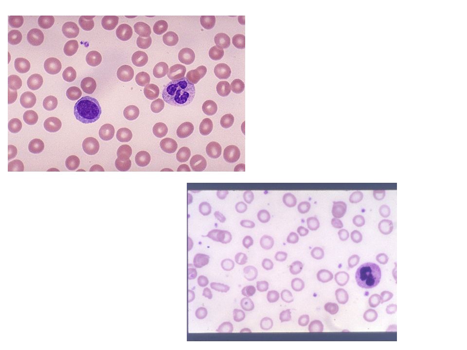
Laborvizsgálatok Most often incidental finding CBC and smear –Hb, WBC, plt –MCV Reticulocyte count Depending on suspected process –Ferritin and iron studies, folate and B12, LDH, bili, Coombs, osmotic fragility, sickle screen, electrophoresis, bone marrow aspiration, renal function, liver function, etc

22
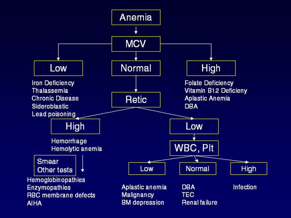
Morphológiai osztályozás Vvt nagyság szerint –Microcyter –Macrocyter –Normocyter

23
Microcyter anaemia Low MCV Small cells Etiologies –Iron-deficiency –Thalassemia –Chronic disease –Lead poisoning

24
Macrocyter anaemia High MCV Big cells Etiologies –Folic acid and B12 deficiency –Hypothyroidism –Chronic liver disease –Aplastic anemias

25
Normocyter Anaemia Normal MCV Production, destruction or loss Must look at reticulocyte count –Young RBC –Determines adequacy of bone marrow response

26
Normocyter anaemia High retic count –Blood loss –Hemolysis Low retic or normal count –Aplastic anemia –Leukemia –Chronic disease –TEC –Congenital hypoplastic syndromes

27
Vashiányos anaemia Epidemiology –Most common heme abnormality of childhood –Most common nutritional deficiency worldwide –500 million to 2 billion people are iron- deficient according to WHO 80% of world’s population

28
Vas metabolismus Body iron content: 2 to 6 g (2 g in adult female, 6 g in adult male) –1.5-2 g in Hb –0.5-1 g bound to enzymes, transferrin (protein that carries iron), in storage form (hemosiderin and ferritin) –The rest in myoglobin –At birth, most term infants have 75 mg of elemental iron per kilogram of body weight, found primarily as hemoglobin (75%), but also as storage (15%) and tissue protein iron (10%). Most iron is recycled Gut absorption depends on: –Epo production –Body iron stores –Bioavailability of dietary iron

29
Vas források Bioavailability factors –Fish, poultry, meat Iron 30% bioavailable –Vegetables Iron 10% bioavailable Absorbtion factors –Vitamin C increases absorbtion –Phytates (bran, oats, rye, fiber) and tea decrease absorbtion
 and tea decrease absorbtion")
30
Vasszükséglet Érett újszülött becsült napi vasszükséglete: 1 mg/kg A vasraktárak több mint 80%-a az utolsó trimesterben raktározódik. A koraszülöttek vasigénye ezért posztnatalisan fokozott. Koraszülöttek napi vasigénye: –1500 – 2500 gr – 2mg/kg/nap –< 1500 g – 4 mg/kg/nap Koraszülöttek igénye erythropoietin kezelés esetében - 6 mg/kg/nap

31
Vas felszívódás Generally 10% of dietrary iron is absobed Greater than 50% of iron from human milk is absorbed compared with typically less than 12% of iron from cow milk-derived formula

32
Rizikócsoportok Babies –Newborn body contains 75mg/kg –Infants triple blood volume in 1 st year –Each kg gain requires increase of 35 to 45 mg body iron –Term babies usually iron replete for 5-6 months, then need iron-fortified foods Iron recycled in first 2 to 3 months –Pre term at greater risk

33
Rizikócsoportok Toddlers –Too much cow’s milk!!! Maximum 16 oz/day Interferes with absorption from other food Colitis Decreased appetite for food Teenagers –Increased requirement due to growth spurt –Menstrual loss

34
Vashiány okai Newborn factors –LBW, perinatal hemorrhage, prematurity Dietary deficiencies –Insufficient intake, poor iron bioavailability Early cow’s milk exposure –Excessive cow’s milk intake Blood loss Iron deficiency itself! –Blunting of intestinal villi leads to increased blood loss

35
Lab eltérések Bone marrow hemosiderin first disappears –Most reliable indicator of tissue stores RDW earliest sign on blood work Ferritin Iron, TIBC Hb Smear –microcytosis and hypochromia Retic normal or moderately increased –Insufficient response

37
Vashiány hosszútávú következményei Has been linked to ADHD and breath-holding spells –Although not well substantiated Exercise intolerance –Study done in teenagers Neurological impairment –More school difficulties, especially math and memory skills Increases lead absorption –Leading to more cognitive abnormalities

38
Szűrés 9 hónaposan ha rizikócsoportba tartozik –Canadian Task force ajánlása Magas rizikó –Kora- vagy sorvadt újsz. –Magas prevalenciájú közösségekben –Alacsony szociális helyzet –Speciális igényű páciensek Szűrés megfontolandó –Kisdedeknél hiányos diétával –Serdülők

39
Prevenció Anyatejes csecsemő –> 2 adag vasban gazdagított cerealia 6 hónapos kortól Tápszeres csecsemő –Vassal szupplementált formula alkalmazása Koraszülött anyatejes –Vas 1 to 2 mg/kg 1-6-12 hónapig 12 hónapos korig nincs tehéntej Tehéntej fogyasztásának limitálása kisdedeknél –< 500 ml 1-5 év között

40
Kezelés Diéta módosítás –Decrease cow’s milk –Increase iron-rich foods Vas terápia –4 to 6 mg/kg of elemental iron Increased absorption with Vit C

41
Kezelés Parenteral iron –2-3% anaphylaxis –No advantage PRBC transfusion –RARELY necessary –Only if hemodynamically unstable –3 to 5 cc/kg at a time, watch for CHF

42
Kezelésre adott válasz 12-24 hours –Intracellular replaced –Subjective improvement –Increased appetite 36-48 hours –Bone marrow response –Erythroid hyperplasia 48 to 72 hours –Retics increased, peak at around 5 to 7 days

43
Kezelésre adott válasz 4 to 30 days –Increased Hemoglobin 1 to 3 months –Repletion of iron stores Treat for a total of at least 3 months

44
Sikertelen kezelés Rossz compliance –10% GI mellékhatások –Rossz íz Folyamatos vérvesztés Rossz diagzózis

46
Különböző anaemiák jellemzői Parameterek, indexek Apl. Anaemia Folate, B12 def. Fe def.HemolysisVérvesztés MCV Hgb conc RBC Hgb conc Rets Se Bil

47
Mikor forduljunk gyermek hematológushoz? Neutropaenia és/vagy thrombocytopaenia Jelentős adenopathia/organomegalia Haemolysis gyanuja Hemodynamikai instabilitás és/vagy HCT < 20% Thalassemia major vagy sarlósejtes anaemia gyanuja Sikertelen vas szubsztitució esetén

48
Hereditary spherocytosis Prevalence: 1/5000 Etiology: Autosomal dominant, 25% have no previous family history Most common molecular defects: spectrin, ankyrin

49
Clinical manifestations Hyperbilirubinemia in the neonate Some children stay symptomeless until adulthood, others have recurrent hemolytic crisis After infancy the spleen is enlarged Gallstone Icterus

50
Treatment Transfusion Splenectomy

51
Diamond-Blackfan anemia Diamond-Blackfan anemia (DBA), also known as Blackfan–Diamond anemia and Inherited erythroblastopenia, is a congenital erythroid aplasia that usually presents in infancy. DBA patients have low red blood cell counts (anemia). The rest of their blood cells (the platelets and the white blood cells) are normal. This is in contrast to Schwachman-Diamond syndrome, in which the bone marrow defect results primarily in neutropenia, and Fanconi anemia, where all cell lines are affected resulting in pancytopenia.congenitalerythroidaplasiainfancy red blood cellanemiaplateletswhite blood cellsSchwachman-Diamond syndromebone marrowneutropeniaFanconi anemiapancytopenia
. The rest of their blood cells (the platelets and the white blood cells) are normal. This is in contrast to Schwachman-Diamond syndrome, in which the bone marrow defect results primarily in neutropenia, and Fanconi anemia, where all cell lines are affected resulting in pancytopenia.congenitalerythroidaplasiainfancy red blood cellanemiaplateletswhite blood cellsSchwachman-Diamond syndromebone marrowneutropeniaFanconi anemiapancytopenia.")
52
Clinical picture Diamond-Blackfan anemia is characterized by anemia (low red blood cell counts) with decreased erythroid in the bone marrow. This usually develops during the neonatal period. About 47% of affected individuals also have a variety of congenital abnormalities, including craniofacial malformations, thumb or upper limb abnormalities, cardiac defects, urogenital malformations, and cleft palate. Low birth weight and generalized growth delay are sometimes observed. DBA patients have a modest risk of developing leukemia and other malignanciesanemiared blood cellbone marrow neonatalcongenital craniofacialurogenitalcleft palateleukemia

53
Diagnosis Typically, a diagnosis of DBA is made through a simple blood count and a bone marrow biopsy.blood countbone marrow biopsy A diagnosis of DBA is made on the basis of anemia, low reticulocyte (immature red blood cells) counts, and diminished erythroid precursors in bone marrow. Features that support a diagnosis of DBA include the presence of congenital abnormalities, macrocytosis, elevated fetal hemoglobin, and elevated adenosine deaminase levels in red blood cells.reticulocyte macrocytosisfetal hemoglobinadenosine deaminase Most patients are diagnosed in the first two years of life. However, some mildly affected individuals only receive attention after a more severely affected family member is identified. About 20-25% of DBA patients may be identified with a genetic test for mutations in the RPS19 gene.(19q13.2)genetic testRPS19
genetic testRPS19.")
54
Treatment Corticosteroid Transfusion Bown marrow transplantation

55
Transient erythroblastopenia of childhood (TEC) Acquired erythroid bone marrow failure –Unknown etiology 18 mos to 2 yrs Often follows viral illness Child otherwise healthy Resolves spontaneously –Weeks to months
 Acquired erythroid bone marrow failure –Unknown etiology 18 mos to 2 yrs Often follows viral illness Child otherwise healthy Resolves spontaneously –Weeks to months")
56
TEC Lab findings –Normocytic anemia (Hb 50-70, sometimes as low as 20) –Low retic count –No evidence of hemolysis –Other cell lines unaffected Treatment –Supportive –Transfusion if symptomatic
 –Low retic count –No evidence of hemolysis –Other cell lines unaffected Treatment –Supportive –Transfusion if symptomatic")
57
Hemoglobinopathies Thalassemia –Decreased or absence of production of one or more globin chains Alpha, beta, and variants Sickle cell disease –Structural defect of beta-globin chain

58
Thalassemia Epidemiology –Prevalent in certain populations Africa, Middle East, Asia, and Mediterranean population Pathogenesis –Decreased or absent synthesis of one or more globin chains Imbalance in number of chains –Precipitation of unstable Hb Hemolysis occurs

59
Alpha-Thalassemia Common in Asian and black populations Phenotype depends on number of deletions –1-gene deletion Silent carrier, no anemia –2-gene deletion (trait) Mild hypochromic, microcytic anemia –3-gene deletion (Hemoglobin H) Severe anemia Hb Bart (gamma globin tetramers) –4-gene deletion Incompatible with life Hb Bart
 Mild hypochromic, microcytic anemia –3-gene deletion (Hemoglobin H) Severe anemia Hb Bart (gamma globin tetramers) –4-gene deletion Incompatible with life Hb Bart")
60
Beta-thalassemia Mediterranean or Southeast Asian origin Phenotype depends on number and type of mutations –Minor Microcytic anemia –Intermedia Moderate to severe anemia –Major Severe anemia, transfusion dependant

61
Diagnosis Family history, ethnic origin CBC –Microcytic anemia Normal or increased ferritin Hb electrophoresis –Decreased HbA, increased HbF –Abnormal Hemoglobins Gene studies

62
Treatment Alpha trait –None –Counseling Severe anemia –Transfusion –Watch for iron overload –Chelation therapy –Splenectomy

63
Sickle Cell Disease Defect of beta-globin chain –Amino acid substitution (valine for glutamine) Prevalent in certain populations –African, Caribbean, Middle Eastern, Indian, Mediterranean populations Hb susceptible to deoxygenation, acidosis, temperature, dehydration –RBC distorted into sickle shape –Results in tissue ischemia and infarction –Shortened RBC survival
 Prevalent in certain populations –African, Caribbean, Middle Eastern, Indian, Mediterranean populations Hb susceptible to deoxygenation, acidosis, temperature, dehydration –RBC distorted into sickle shape –Results in tissue ischemia and infarction –Shortened RBC survival")
64
Sickle Cell Disease Manifestations (only in disease, not if trait) –Bony crisis –Chest crisis –Strokes –Splenic sequestration –Aplastic anemia Susceptibility to infections; autosplenectomize –Encapsulated organisms Need penicillin prophylaxis
 –Bony crisis –Chest crisis –Strokes –Splenic sequestration –Aplastic anemia Susceptibility to infections; autosplenectomize –Encapsulated organisms Need penicillin prophylaxis")
65
Treatment Pain control –NSAIDs, opiates Hydration Antibiotics if febrile Transfusions Exchange transfusions Hydroxyurea

Hasonló előadás


![ELÉG MAGYAR MÉRNÖKÜNK VAN A GAZDASÁGI ÉS INNOVÁCIÓS KITÖRÉSHEZ? TAMÁS PÁL [MTA Szociológiai Kutatóintézet, Budapest]](/9/2218527/big_thumb.jpg "ELÉG MAGYAR MÉRNÖKÜNK VAN A GAZDASÁGI ÉS INNOVÁCIÓS KITÖRÉSHEZ? TAMÁS PÁL [MTA Szociológiai Kutatóintézet, Budapest]>")