Előadást letölteni
Az előadás letöltése folymat van. Kérjük, várjon

1
Autoimmunitás/Autoimmunitás kialakulásának okai

2
Populáció ~5% szenved autoimmun betegségben

4
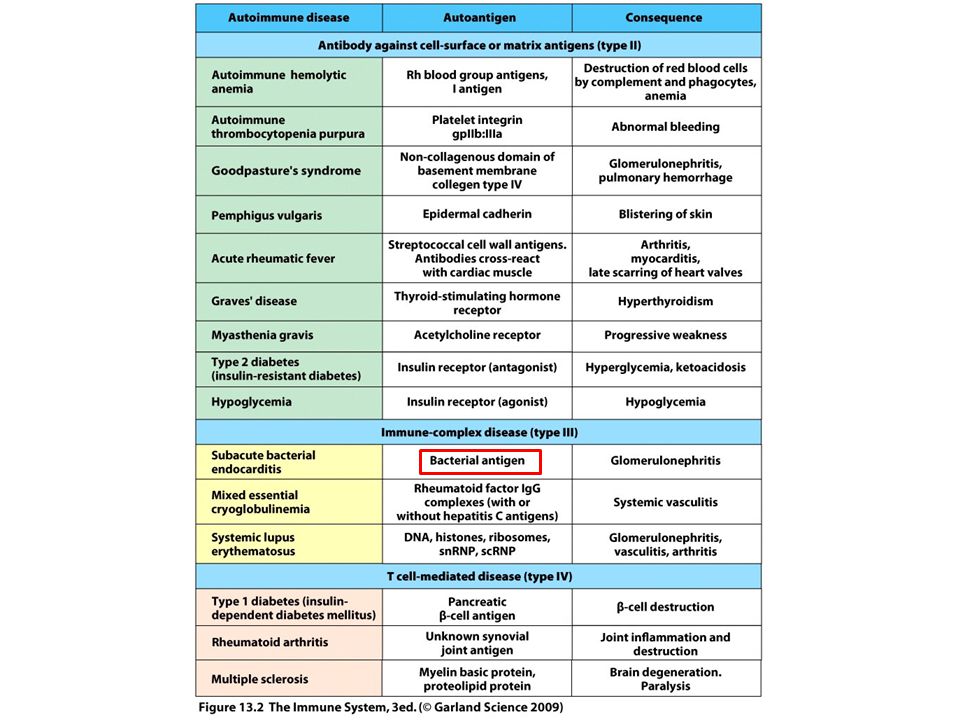
TÚLÉRZÉKENYSÉGI REAKCIÓK ÁTTEKINTÉSE/Autoimmunitás? I. típusú
„azonnali” II. típusú III. típusú IV. típusú „késői” Ellenanyag mediált T sejt mediált specifikus IgE sejtfelszíni antigénnel specifikusan reagáló ellenanyag, IgG aspecifikusan lerakódó, szolubilis immunkomplex MHC függő T sejt aktiváció hízósejtekből felszabaduló mediátor anyagok FcR mediált, gyuladás, sejtfunkció gátlás FcR mediált, komplement aktiváció, gyulladás citokinek, citotoxicitás „Klasszikus allergia” újszülöttkori hemolitikus anémia, (penicillin?) érzékenység, M. gravis szérumbetegség, SLE kontakt drematitisz M. Gravis – izomsejteken levő acethilcolin receptorokhoz kötődő IgG ellenanyagok blokkolják a jelátvitelt az idegvégződésekről az izomra Igen gyakran autoimmun betegségek együttjárói
 érzékenység, M. gravis. szérumbetegség, SLE. kontakt drematitisz. M. Gravis – izomsejteken levő acethilcolin receptorokhoz kötődő IgG ellenanyagok blokkolják a jelátvitelt az idegvégződésekről az izomra. Igen gyakran autoimmun betegségek együttjárói.")
6
Tolerancia

7
Molecular Mechanisms of Autoimmunity
How is autoimmunity induced? What could go wrong here? 1-immunologic factor 2-genetic f. 3-enviroment f. 7

8
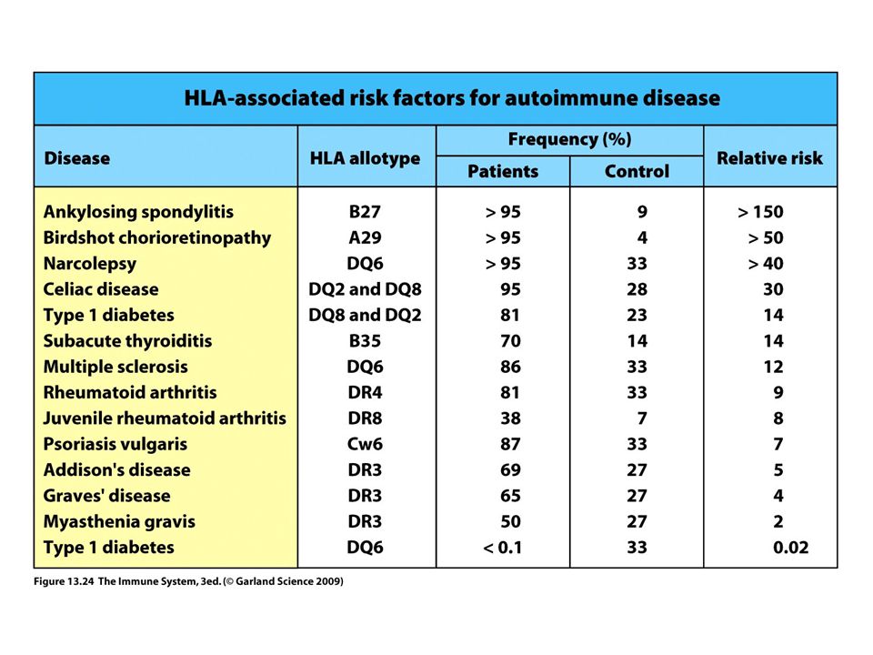
Genetikai tényezők Much recent attention has focused on the role of T cells in autoimmunity, for two main reasons. First, helper T cells are the key regulators of all immune responses to proteins, and most self antigens implicated in autoimmune diseases are proteins. Second, several autoimmune diseases are genetically linked to the MHC (the HLA complex in humans), and the function of MHC molecules is to present peptide antigens to T cells.
, and the function of MHC molecules is to present peptide antigens to T cells.")
10
Protein tyrosine phosphatase; role in T and B cell receptors signaling
Chromosomal Region Gene of Interest Function Diseases Genes Involved in Immune Regulation 1p13 PTPN22 Protein tyrosine phosphatase; role in T and B cell receptors signaling RA, T1D, IBD 1p12 CD2/CD58 Costimulation of T cells RA, MS 1p31 IL23R Component of IL-23 receptor; role in generation and maintenance of TH17 cells IBD, PS, AS 1q32 IL10 Downregulates expression of costimulators, MHC molecules, IL-12 in dendritic cells; inhibits TH1 responses IBD, SLE, T1D 2q33 CTLA4 Inhibitory receptor of T cells, effector molecule of regulatory T cells T1D, RA 4q26 IL2/IL21 Growth and differentiation factors for T cells; IL-2 is involved in maintenance of functional Tregs IBD, CeD, RA, T1D, MS 5q33 IL12B p40 subunit of IL-12 (TH1-inducing cytokine) and IL-23 (TH17-inducing cytokine) IBD, PS 8p23 BLK B lymphocyte tyrosine kinase, involved in B cell activation SLE, RA 10p15 IL2RA IL-2 receptor α chain (CD25); role in T cell activation and maintenance of regulatory T cells MS, T1D Genes Involved in Responses to Microbes 16q12 NOD2 Cytoplasmic sensor of bacteria IBD 2q37 ATG16 Autophagy (destruction of microbes, maintenance of epithelial cell integrity) 7q32, 2q24 IRF5, IFIH1 Type I interferon responses to viruses SLE Table Selected Non-HLA Genetic Associations with Autoimmune Diseases Insulin polimorf vsz befolyásolja, hogy megjelenik-e a tímuszban AS, ankylosing spondylitis; CeD, celiac diseases; IBD, inflammatory bowel disease; MS, multiple sclerosis; PS, psoriasis; RA, rheumatoid arthritis; SLE, systemic lupus erythematosus; T1D, type 1 diabetes. Data from Zenewicz L, C Abraham, RA Flavell, and J Cho. Unraveling the genetics of autoimmunity. Cell 140: , 2010, with permission of the publisher.
 and IL-23 (TH17-inducing cytokine) IBD, PS. 8p23. BLK. B lymphocyte tyrosine kinase, involved in B cell activation. SLE, RA. 10p15. IL2RA. IL-2 receptor α chain (CD25); role in T cell activation and maintenance of regulatory T cells. MS, T1D. Genes Involved in Responses to Microbes. 16q12. NOD2. Cytoplasmic sensor of bacteria. IBD. 2q37. ATG16. Autophagy (destruction of microbes, maintenance of epithelial cell integrity) 7q32, 2q24. IRF5, IFIH1. Type I interferon responses to viruses. SLE. Table Selected Non-HLA Genetic Associations with Autoimmune Diseases. Insulin polimorf vsz befolyásolja, hogy megjelenik-e a tímuszban. AS, ankylosing spondylitis; CeD, celiac diseases; IBD, inflammatory bowel disease; MS, multiple sclerosis; PS, psoriasis; RA, rheumatoid arthritis; SLE, systemic lupus erythematosus; T1D, type 1 diabetes. Data from Zenewicz L, C Abraham, RA Flavell, and J Cho. Unraveling the genetics of autoimmunity. Cell 140: , 2010, with permission of the publisher.")
11
Phenotype of Mutant or Knockout Mouse
Table Examples of Single-Gene Mutations That Cause Autoimmune Diseases Gene Phenotype of Mutant or Knockout Mouse Mechanism of Failure of Tolerance Human Disease? AIRE Destruction of endocrine organs by antibodies, lymphocytes Failure of central tolerance Autoimmune polyendocrine syndrome (APS) C4 SLE Defective clearance of immune complexes; failure of B cell tolerance? CTLA-4 Lymphoproliferation; T cell infiltrates in multiple organs, especially heart; lethal by 3-4 weeks Failure of anergy in CD4+ T cells; defective function of regulatory T cells CTLA-4 polymorphisms associated with several autoimmune diseases Fas/FasL Anti-DNA and other autoantibodies; immune complex nephritis; arthritis; lymphoproliferation Defective deletion of anergic self-reactive B cells; reduced deletion of mature CD4+ T cells Autoimmune lymphoproliferative syndrome (ALPS) FoxP3 Multiorgan lymphocytic infiltrates, wasting Deficiency of functional regulatory T cells IPEX IL-2, IL-2Rα/β Inflammatory bowel disease; anti-erythrocyte and anti-DNA autoantibodies Defective development, survival, or function of regulatory T cells None known SHP-1 Multiple autoantibodies Failure of negative regulation of B cells AIRE, autoimmune regulator gene; IL-2, interleukin-2; IPEX, immune dysregulation, polyendocrinopathy, enteropathy, X-linked syndrome; SHP-1, SH2-containing phosphatase 1; SLE, systemic lupus erythematosus.
 C4. SLE. Defective clearance of immune complexes; failure of B cell tolerance CTLA-4. Lymphoproliferation; T cell infiltrates in multiple organs, especially heart; lethal by 3-4 weeks. Failure of anergy in CD4+ T cells; defective function of regulatory T cells. CTLA-4 polymorphisms associated with several autoimmune diseases. Fas/FasL. Anti-DNA and other autoantibodies; immune complex nephritis; arthritis; lymphoproliferation. Defective deletion of anergic self-reactive B cells; reduced deletion of mature CD4+ T cells. Autoimmune lymphoproliferative syndrome (ALPS) FoxP3. Multiorgan lymphocytic infiltrates, wasting. Deficiency of functional regulatory T cells. IPEX. IL-2, IL-2Rα/β. Inflammatory bowel disease; anti-erythrocyte and anti-DNA autoantibodies. Defective development, survival, or function of regulatory T cells. None known. SHP-1. Multiple autoantibodies. Failure of negative regulation of B cells. AIRE, autoimmune regulator gene; IL-2, interleukin-2; IPEX, immune dysregulation, polyendocrinopathy, enteropathy, X-linked syndrome; SHP-1, SH2-containing phosphatase 1; SLE, systemic lupus erythematosus.")
12
Immunológiai tényezők
Defects in deletion (negative selection) of T or B cells or receptor editing in B cells during the maturation of these cells in the generative lymphoid organs Defective numbers and functions of regulatory T lymphocytes Defective apoptosis of mature self-reactive lymphocytes Inadequate function of inhibitory receptors Activation of APCs, which overcomes regulatory mechanisms and results in excessive T cell activation
 of T or B cells or receptor editing in B cells during the maturation of these cells in the generative lymphoid organs. Defective numbers and functions of regulatory T lymphocytes. Defective apoptosis of mature self-reactive lymphocytes. Inadequate function of inhibitory receptors. Activation of APCs, which overcomes regulatory mechanisms and results in excessive T cell activation.")
13
Several ways in which infectious agents could break self tolerance

14
Adjuvans+saját antigén

15
Ubiquitináció-degradáció
Foszfatáz Ubiquitináció-degradáció +Citokin környezet CTLA-4 nagyobb affinitású, mint a CD28 mice lacking CTLA-4 develop uncontrolled lymphocyte activation with massively enlarged lymph nodes and spleen and fatal multiorgan lymphocytic infiltrates suggestive of systemic autoimmunity. The autoimmune disorders in PD-1 knockout mice are less severe than in CTLA-4 knockouts. It has been postulated that CTLA-4 functions mainly to control initial T cell activation in lymphoid organs whereas PD-1 is more important for limiting responses of differentiated effector cells in peripheral tissues.

16
Figure 7-22 Role of the ubiquitin ligase Cbl-b in terminating T cell responses. Cbl-b is recruited to the TCR complex, where it facilitates the monoubiquitination of CD3, ZAP-70, and other proteins of the TCR complex. These proteins are targeted for proteolytic degradation in lysosomes and other organelles (not shown). . Recruitment of Cbl-b to the TCR complex and associated adaptor proteins leads to the monoubiquitination, endocytosis, and lysosomal degradation of the TCR complex, and this may be a mechanism for the attenuation of TCR signaling (Fig. 7-22). CD28 signals block the inhibitory activity of Cbl-b, and this is one mechanism by which costimulation augments TCR signals. In knockout mice lacking Cbl-b, the T cells respond to antigen even without CD28-mediated costimulation and produce abnormally high amounts of IL-2. These mice develop autoimmunity as a result of the enhanced activation of their T cells.
. CD28 signals block the inhibitory activity of Cbl-b, and this is one mechanism by which costimulation augments TCR signals. In knockout mice lacking Cbl-b, the T cells respond to antigen even without CD28-mediated costimulation and produce abnormally high amounts of IL-2. These mice develop autoimmunity as a result of the enhanced activation of their T cells.")
17
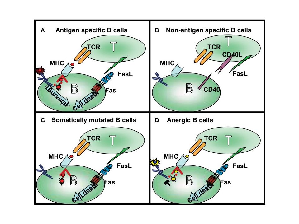
Peripheral B-cell anergy

21
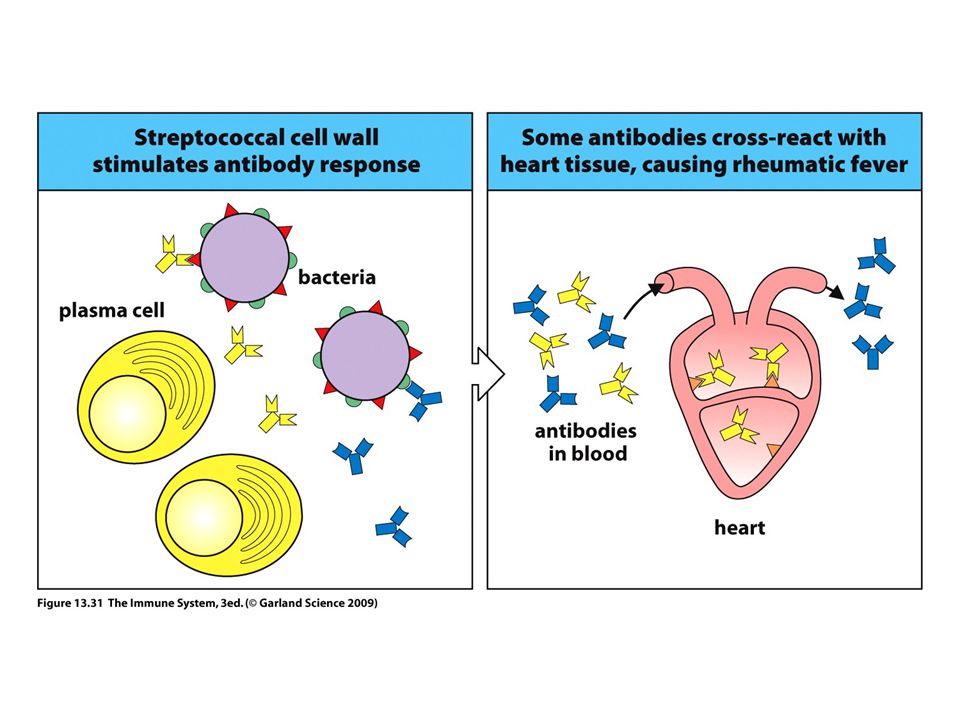
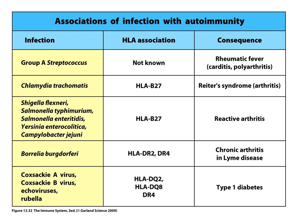
Patogén specifikus– kereszt reaktivitás

22
Cross-Reactivity 22

23
Patogén független Pl. fokozott MHC expresszió

24
epitóp terjedés

25
Intramolekuláris epitóp terjedés

26
Szuperantigének (pl. bakteriális toxinok) az MHC II molekulákhoz és a T-sejt receptorokhoz kívülről ( nem a specifikus antigén-kötő helyen) kapcsolódnak, így egyszerre nagy számú nem specikus T-sejt proliferációját idézhetik elő.
 az MHC II molekulákhoz és a T-sejt receptorokhoz kívülről ( nem a specifikus antigén-kötő helyen) kapcsolódnak, így egyszerre nagy számú nem specikus T-sejt proliferációját idézhetik elő..")
27
4. Polyclonal Activation Hypothesis
B-cell mitogens, e.g. LPS, EBV Bact. Superag. TCR (V) -self or Th BUT Limited specificity, e.g. thyroiditis Clonally restricted e.g. -DNA in SLE 27
 -self or Th. BUT. Limited specificity, e.g. thyroiditis. Clonally restricted e.g. -DNA in SLE. 27.")
28
Danger Theory Anti-self B & T-cells always present.
AIR is due to release of “danger signals.” Response to tissue damage, necrosis or cell distress, e.g. infection or injury. BUT AIR can occur without tissue damage, e.g. immunisn. with self-ag; Tx; genetic defects. 28

29
Summary Self reactive B-cells & T-cells are normally present but anergic. Several factors can induce an AIR:- Genetic Tissue damage & release of cryptic ag. Somatic mutation in Ig V-genes Ag mimicry Tr defects Danger signals 29

30
BAFF– B cell activating faktor (B-sejtek fitnesz faktora)
. A BAFF konstitutív expressziója kell az érett B-sejtek hosszútávú túléléséhez. Nincs BAFF nincs érett B-sejt, overexpressszió megnövekedett B-sejt szám. A BAFF normál körülmények között limitáltan termelődik. BAFFR van szerepe BCMA, TACI-nak nincs.

31
BAFF A B-sejtek versenyeznek a limitált szabad BAFF-ért
Több BAFF jelenléte esetén több B-sejt maradhat életben A B-sejtek versenyeznek a limitált szabad BAFF-ért Michael P., 2009, Nat. Rev. Immunol.

Hasonló előadás