Előadást letölteni
Az előadás letöltése folymat van. Kérjük, várjon

1
B-sejt típusok B1-sejtek Antigén nélkül is ellenanyagot termelnek Az ellenanyag ált. IgM, polispecifikus, nem/ nem csak fehérje antigének A VDJ átrendeződés nem véletlenszerű, TdT nem expresszálódik bennük—minden egyedben megjelennek, ugyanazokat az antigéneket ismerik fel Önfenntartó, hosszú életű sejt populáció CLL

2
Figure 7-41

3
(HOL ÉS HOGYAN TÖRTÉNIK?)
B-SEJT AKTIVÁCIÓ (HOL ÉS HOGYAN TÖRTÉNIK?)
")
4
FDC-ek felszínén ‘tárolódik’.
HEV-high endothelial venule Speciális endothél sejtek, csak a nyirokcsomóban található, mely elősegíti a limfociták (B és T) kilépését a keringésből a nyirokcsomókba) Az antigén a szövetek közül a nyirokcsomóba kerül (Főként a DC-ek által), ahol a FDC-ek felszínén ‘tárolódik’.
 kilépését a keringésből a nyirokcsomókba) Az antigén a szövetek közül a nyirokcsomóba kerül (Főként a DC-ek által), ahol a. FDC-ek felszínén ‘tárolódik’.")
5
Immunkomplex (1)antigén-(2)az ezt felismerő ellenanyag és (3)komplement fehérjék komplexe Komplement fehérjék

6
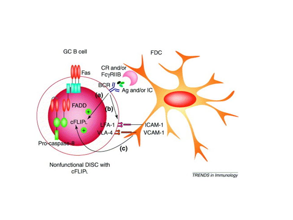
A B sejtek az FDC felszínén ismerik fel az antigént
! Az antigén a follikuláris dendritikus sejtek (FDC) felszínéhez kötődik (Az FDC felszínén állandóan jelenlevő Fc és komplement recepotrokon keresztül) Az FDC-ek immun komplexeket kötnek (Ag-Ab) Az antigén ingert követően 12 hónappal is kimutatható az FDC-ek felszínén Egyetlen sejt sokféle antigént köthet Develop from fibroblast-like cells FDC fugg a TNFa, Lta, LTb jelenlététől A B sejtek az FDC felszínén ismerik fel az antigént 6
 felszínéhez kötődik (Az FDC felszínén állandóan jelenlevő Fc és komplement recepotrokon keresztül) Az FDC-ek immun komplexeket kötnek (Ag-Ab) Az antigén ingert követően 12 hónappal is kimutatható az FDC-ek felszínén. Egyetlen sejt sokféle antigént köthet. Develop from fibroblast-like cells. FDC fugg a TNFa, Lta, LTb jelenlététől. A B sejtek az FDC felszínén ismerik fel az antigént. 6.")
7
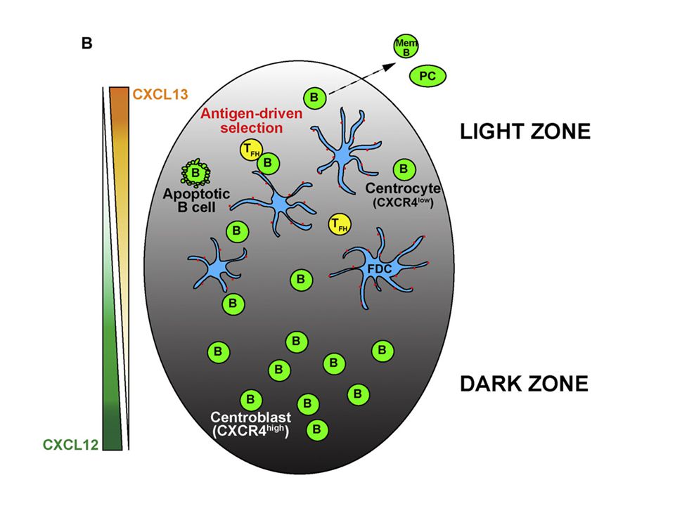
A germinális centrum (csíra központ) szerkezete
Szomatikus hypermutáció LZ FDC DZ LZ: világos zóna DZ: sötét zóna FDC: follikuláris dendritikus sejt

9
A B-sejtek ‘versengenek’ az antigénért
! A B-sejtek ‘versengenek’ az antigénért A nagy affinitású B-sejtek nagyobb valószínűséggel jutnak antigénhez

10
! A B-sejtek antigén prezentációja
Az antigén megkötése a B-sejt receptor által kiváltja a B-sejt aktivációt Az antigén felvételét (endocitózisát) és ezáltal MHCII általi prezentációját A prezentáció következtében ugyanazat az antigént felismerő B és T-sejtek válogatódnak ki. A T-sejtek segítő és túlélő jeleket biztosítanak a B-sejtek számára Antigén feldolgozás és bemutatás A T-sejtek antigén felismerése
 és ezáltal MHCII általi prezentációját. A prezentáció következtében ugyanazat az antigént felismerő B és T-sejtek válogatódnak ki. A T-sejtek segítő és túlélő jeleket biztosítanak a B-sejtek számára. Antigén feldolgozás és bemutatás. A T-sejtek antigén felismerése.")
11
+++ Antigén prezentáció A poliszaharidok nem prezentálódnak!
A prezentáció következtében ugyanazat az antigént felismerő B és T-sejtek válogatódnak ki +++ A poliszaharidok nem prezentálódnak!

12
2. A B-sejtek ‘versengenek’ A T-sejtek segítő szignáljáért
! 2. A B-sejtek ‘versengenek’ A T-sejtek segítő szignáljáért A nagy affinitású B-sejtek nagyobb valószínűséggel prezentálnak antigént A B-sejtek versengenek

13
A sikeres B-sejtek a T-sejttel való találkozás után újra kezdik a ciklust
Osztódnak Szomatikus mutációs lépéseken esnek át. versengenek az antigénért, 4. Újra versengenek aT-sejtek segítő szignáljáért. A ciklusok során a szomatikus mutációnak és a szelekciónak köszönhetően a sejtfelszíni ellenanyag affinitása nő—affinitás érés A ciklus ismétlődik vagy a B-sejtek kilépnek a ciklusból. Plazma vagy memória sejtté differenciálódnak

14
Azoknak a B-sejtek van a legnagyobb esélye a T-sejt ‘helpre, amik ugyanazt az antigént ismerik fel, mutatják be, mint amit a DC-k) B-sejtek bemutatják a T-sejteknek az antigént.

15
PROLIFERÁCIÓ/DIFFERENCIÁCIÓ
A B-sejt aktiváció fő lépései AKTIVÁCIÓ PROLIFERÁCIÓ/DIFFERENCIÁCIÓ FELISMERÉS Ea termelés Klonális szaporodás Segítő T Izotípus váltás Szomatikus mutáció-Affinitás érés Memória B sejt A sikeres –szelekción átesett- B-sejtek plazmasejtekké vagy memória sejtekké differenciálódnak

16
A B-sejtek mozgása a GC-ban a sötét és a világos zóna között
The light zone FDCs are identified by in vivo deposited PE immune complexes (red). Colored lines show tracks of GC B cells that moved within the light or dark zones or that crossed between zones, as indicated, during a 30 minute imaging session. The gridlines are separated by 20µm. Cell tracks were manually classified as being in the light or dark zones if the entire track stayed within the PE+ or PE− region, respectively, during the imaging session. Cell tracks were manually classified as traveling between zones if the tracks originated in one zone, crossed an approximately 20 µm border between the zones, then entered at least 10 µm into the other zone, and stayed in that zone for the duration of the analysis. Some tracks could not be definitively classified by these criteria, for example tracks that were too close to the border between the light and dark zones. From Allen et al., Science 315: 528 (2007).
. Colored lines show tracks of GC B cells that moved within the light or dark zones or that crossed between zones, as indicated, during a 30 minute imaging session. The gridlines are separated by 20µm. Cell tracks were manually classified as being in the light or dark zones if the entire track stayed within the PE+ or PE− region, respectively, during the imaging session. Cell tracks were manually classified as traveling between zones if the tracks originated in one zone, crossed an approximately 20 µm border between the zones, then entered at least 10 µm into the other zone, and stayed in that zone for the duration of the analysis. Some tracks could not be definitively classified by these criteria, for example tracks that were too close to the border between the light and dark zones. From Allen et al., Science 315: 528 (2007).")
17
Hogyan jut az antigén a GC-ba?
Hogyan jut az antigén az FDC-k felszínére? Az ellenanyagtermelés (IgG) GC-ot igényel, milyen immunkomplex tapad a FDC felszínre? Az FDC hosszú ideig tartja az antigént, a B-sejt prezentál? Versengés az antigénért/versengés a T-sejt segítő szignálért?
 GC-ot igényel, milyen immunkomplex tapad a FDC felszínre Az FDC hosszú ideig tartja az antigént, a B-sejt prezentál Versengés az antigénért/versengés a T-sejt segítő szignálért")
18
Small antigens, not bigger than 70KDa
Conduits mediate transport of low-molecular-weight antigen to lymph node follicles. Immunity Feb Roozendaal R, Mempel TR, Pitcher LA, Gonzalez SF, Verschoor A, Mebius RE, von Andrian UH, Carroll MC. The LN is supplied with lymphatic fluid containing antigens and chemokines (shown in red and green, respectively) through the afferent lymph vessel. On arrival, CD169+ macrophages lining the SCS prevent free diffusion of lymphatic fluid to the LN interior. Larger antigens (diamonds) are accumulated by SCS macrophages and can be directly presented to antigen-specific B cells (the B cell receptor is shown in red) in the follicle. In addition, larger antigens can be transported by nonspecific B cells to FDCs in a complement-dependent manner. This transport may allow FDCs to mediate antigen presentation to specific naive B cells. In contrast, smaller (less than 70 kDa) antigens and chemokines (circles) can access antigen-specific B cells in the follicle either by direct diffusion from pores in the SCS or via the follicular conduit system. These follicular conduits extend from the SCS and are comprised of bundles of collagen fibers ensheathed by fibroreticular cells (FRCs). Because the FRC network is not completely continuous, follicular B cells can extend protrusions through tiny gaps to sample the transported components of lymphatic fluid. Small antigens, not bigger than 70KDa
 through the afferent lymph vessel. On arrival, CD169+ macrophages lining the SCS prevent free diffusion of lymphatic fluid to the LN interior. Larger antigens (diamonds) are accumulated by SCS macrophages. and can be directly presented to antigen-specific B cells (the B cell receptor is shown in red) in the follicle. In addition, larger antigens can be transported. by nonspecific B cells to FDCs in a complement-dependent manner. This transport may allow FDCs to mediate antigen presentation to specific naive B cells. In. contrast, smaller (less than 70 kDa) antigens and chemokines (circles) can access antigen-specific B cells in the follicle either by direct diffusion from pores in the. SCS or via the follicular conduit system. These follicular conduits extend from the SCS and are comprised of bundles of collagen fibers ensheathed by fibroreticular. cells (FRCs). Because the FRC network is not completely continuous, follicular B cells can extend protrusions through tiny gaps to sample the transported. components of lymphatic fluid. Small antigens, not bigger than 70KDa.")
19
Zöld –antigén Kék B-sejt

20
70 Kda-nál kisebb antigének--csatorna
Nagyobb antigének: Kb 1óra múlva makrofágok 4-24 óra B sejtek (naív/MZ B sejt) 24-tól FDC (DCs plasmacytoid dendritic)
 24-tól FDC. (DCs. plasmacytoid dendritic)")
21
DCs FcgRIIB internalized ICs are inefficiently degraded and FcgRIIB-internalized surface recycled ICs promoting antigen-specific B cell activation. Thus, ITAM-mediated signaling in DCs couples efficient degradation of internalized ICs with cellular maturation, Cell surface recycling of internalized antigen permits dendritic cell priming of B cells. Bergtold A, Desai DD, Gavhane A, Clynes R. Immunity Nov;23(5): Whereas macrophages contained high levels of lysosomal proteases and rapidly degraded internalized proteins, dendritic cells (DCs) and B lymphocytes were protease-poor, resulting in a limited capacity for lysosomal degradation. Differential lysosomal proteolysis in antigen-presenting cells determines antigen fate. Science Mar.Delamarre L, Pack M, Chang H, Mellman I, Trombetta ES.
: Whereas macrophages contained high levels of lysosomal proteases and rapidly degraded internalized proteins, dendritic cells (DCs) and B lymphocytes were protease-poor, resulting in a limited capacity for lysosomal degradation. Differential lysosomal proteolysis in antigen-presenting cells determines antigen fate. Science Mar.Delamarre L, Pack M, Chang H, Mellman I, Trombetta ES.")
22
Hogyan jut az antigén a GC-ba?
Hogyan jut az antigén az FDC-k felszínére? Az ellenanyagtermelés (IgG) GC-ot igényel, milyen immunkomplex tapad a FDC felszínre? Az FDC hosszú ideig tartja az antigént, a B-sejt prezentál? Versengés az antigénért/varsengés a T-sejt segítő szignálért?
 GC-ot igényel, milyen immunkomplex tapad a FDC felszínre Az FDC hosszú ideig tartja az antigént, a B-sejt prezentál Versengés az antigénért/varsengés a T-sejt segítő szignálért")
23
Fc receptorok Complement receptorok

24
FDC-k immunkomplexet kötnek ---ellenanyag vagy komplement felismerés
Terrmészetes ellenanyagok?? IgM Natural IgM enhances the immunogenicity of ICs by concentrating IC deposition onto FDCs Curr Opin Immunol Jun;17 Marginal zone B cells in lymphocyte activation and regulation. Lopes-Carvalho T, Foote J, Kearney JF. FcR Taken together, these results suggest that FCMR negatively regulates humoral immune responses against both TI and TD Ags. Mouse IgM Fc receptor, FCMR, promotes B cell development and modulates antigen-driven immune responses. Choi SC, Wang H, Tian L, Murakami Y, Shin DM, Borrego F, Morse HC 3rd, Coligan JE. J Immunol Feb 1; 2. IgG Trapping of IC in spleen and lymph nodes of Fc gamma RII-/- mice was significantly reduced compared with that in wild-type controls. Addition of ICs to cultures of Ag-specific T and B cells elicited pronounced Ab responses only in the presence of FDCs. Fc gamma receptor IIB on follicular dendritic cells regulates the B cell recall response. Qin D, Wu J, Vora KA, Ravetch JV, Szakal AK, Manser T, Tew JG. J Immunol Jun 15

25
A komplement rendszer in vivo szerepe Effektor funkciók
alternatív, lektin és klasszikus út C3 MAC C3a C4a direkt citotoxikus hatás (lízis) C3b C5a gyulladás C3b opszonizáció Különféle immunkomplexek szállítása és fagocitózisa
 C3b. C5a. gyulladás. C3b. opszonizáció. Különféle immunkomplexek szállítása és fagocitózisa.")
26
CR2 Cr2-/- mice demonstrate substantial defects in Ag-specific, T cell-dependent and T cell-independent humoral immune responses (28 –30) that is due to a lack of receptor on both B cells and follicular dendritic cells (31). Cr2/ mice also demonstrate defects in B cell memory (32, 33) and the development of the natural Ab repertoire (34, 35) Studies in mice, which were manipulated to express CR2 only on their B cells or their FDC, have shown that CR2 on B cells plays an important role in initiating a humoral response while FDC CR2 is essential for the maintenance of this response
 that is due to a lack of receptor on both B cells and follicular dendritic cells (31). Cr2/ mice also demonstrate defects in B cell memory (32, 33) and the development of the natural Ab repertoire (34, 35) Studies in mice, which were manipulated to express CR2 only on their B cells or their FDC, have shown that CR2 on B cells plays an important role in initiating a humoral response while FDC CR2 is essential for the maintenance of this response.")
27
Surprisingly, pentameric IgM-containing IC were trapped in spleens of C3-depleted and Cr2-deficient mice. However, the IC were found only in the marginal zone (MZ), associated predominantly with MZ macrophages. IC were also detected in the MZ in normal mice within 1 h after injection The data also provide direct evidence that C3 activation is required for the next phase of localization, in which Ag moves from the MZ to FDC We demonstrate that IgM-IC localize first to the splenic marginal zone (MZ) where the IgM-IC bind MZ B cells in a complement and complement receptor (CR1/2) dependent process. MZ B cells then transport the IgM-IC into the follicle for deposition onto FDCs Marginal zone B cells transport and deposit IgM-containing immune complexes onto follicular dendritic cells. Ferguson AR, Youd ME, Corley RB.
 where the IgM-IC bind MZ B cells in a complement and complement receptor (CR1/2) dependent process. MZ B cells then transport the IgM-IC into the follicle for deposition onto FDCs. Marginal zone B cells transport and deposit IgM-containing immune complexes onto follicular dendritic cells. Ferguson AR, Youd ME, Corley RB.")
28
Figure 4. Pathways for the Recognition of B
Cell Antigen in the Lymph Node Processes depicted in the figure are as follows: (1) Immune complexes (ICs), formed by the deposition of complement proteins (in this illustration, C3d) and IgG on the surface of antigen, bind to complement receptor 3 (CR3) on the surface of subcapsular sinus macrophages (M). (2) Naive B cells transport complement-coated ICs from the subcapsular sinus to FDCs. (3) The ICs are transferred in a complement receptor 2 (CR2)- mediated mechanism from the surface of the B cell to the FDCs. (4) Cognate B cells capture small antigen directly from the surface of FDCs, associated with CR2 receptors (adapted from Gonzalez et al., 2011).
 Immune complexes (ICs), formed by the deposition. of complement proteins (in this illustration, C3d) and IgG on the surface of antigen, bind to. complement receptor 3 (CR3) on the surface of. subcapsular sinus macrophages (M). (2) Naive B. cells transport complement-coated ICs from. the subcapsular sinus to FDCs. (3) The ICs are. transferred in a complement receptor 2 (CR2)- mediated mechanism from the surface of the B cell. to the FDCs. (4) Cognate B cells capture small. antigen directly from the surface of FDCs, associated. with CR2 receptors (adapted from Gonzalez. et al., 2011).")
29
The major role for CD14 is to enhance the sensitivity of the MD-2/TLR4 signaling complex, dropping the binding affinity for LPS to picomolar concentrations The scavenger receptor expressed by phorbol ester-treated rabbit SMCs and fibroblasts bound chemically modified LDL with an order-of-magnitude higher affinity (Kd 5.1 x 10(-10) M)
 M)")
30
High (Kd < 10-9 M); binds IgG1 and IgG3, can bind monomeric IgG
FcR Affinity for Immunoglobulin Cell Distribution Function FcγRI (CD64) High (Kd < 10-9 M); binds IgG1 and IgG3, can bind monomeric IgG Macrophages, neutrophils; also eosinophils Phagocytosis; activation of phagocytes FcγRIIA (CD32) Low (Kd > 10-7 M) Macrophages, neutrophils; eosinophils, platelets Phagocytosis; cell activation (inefficient) FcγRIIB (CD32) B lymphocytes Feedback inhibition of B cells FcγRIIC (CD32) Macrophages, neutrophils, NK cells Phagocytosis, cell activation FcγRIIIA (CD16) Low (Kd > 10-6 M) NK cells Antibody-dependent cell-mediated cytotoxicity FcγRIIIB (CD16) Low (Kd > 10-6 M); GPI-linked protein Neutrophils Phagocytosis (inefficient) FcΕRI High (Kd > M); binds monomeric IgE Mast cells, basophils, eosinophils Cell activation (degranulation) FcΕRII (CD23) B lymphocytes, eosinophils, Langerhans cells Unknown FcαR (CD89) Neutrophils, eosinophils, monocytes Cell activation?
 High (Kd < 10-9 M); binds IgG1 and IgG3, can bind monomeric IgG. Macrophages, neutrophils; also eosinophils. Phagocytosis; activation of phagocytes. FcγRIIA (CD32) Low (Kd > 10-7 M) Macrophages, neutrophils; eosinophils, platelets. Phagocytosis; cell activation (inefficient) FcγRIIB (CD32) B lymphocytes. Feedback inhibition of B cells. FcγRIIC (CD32) Macrophages, neutrophils, NK cells. Phagocytosis, cell activation. FcγRIIIA (CD16) Low (Kd > 10-6 M) NK cells. Antibody-dependent cell-mediated cytotoxicity. FcγRIIIB (CD16) Low (Kd > 10-6 M); GPI-linked protein. Neutrophils. Phagocytosis (inefficient) FcΕRI. High (Kd > M); binds monomeric IgE. Mast cells, basophils, eosinophils. Cell activation (degranulation) FcΕRII (CD23) B lymphocytes, eosinophils, Langerhans cells. Unknown. FcαR (CD89) Neutrophils, eosinophils, monocytes. Cell activation")
31
T Cell Receptor (TCR) Immunoglobulin (Ig) Components α and β chains Heavy and light chains Number of Ig domains One V domain and one C domain in each chain Heavy chain: one V domain, three or four C domains Light chain: one V domain and one C domain Number of CDRs Three in each chain for antigen binding Three in each chain Associated signaling molecules CD3 and ζ Igα and Igβ Affinity for antigen (Kd) M M (secreted Ig) Changes after cellular activation Production of secreted form No Yes Isotope switching Somatic mutations
 M M (secreted Ig) Changes after cellular activation. Production of secreted form. No. Yes. Isotope switching. Somatic mutations.")
32
Hogyan jut az antigén a GC-ba?
Hogyan jut az antigén az FDC-k felszínére? Az ellenanyagtermelés (IgG) GC-ot igényel, milyen immunkomplex tapad a FDC felszínre? Az FDC hosszú ideig tartja az antigént, a B-sejt prezentál? Versengés az antigénért/varsengés a T-sejt segítő szignálért?
 GC-ot igényel, milyen immunkomplex tapad a FDC felszínre Az FDC hosszú ideig tartja az antigént, a B-sejt prezentál Versengés az antigénért/varsengés a T-sejt segítő szignálért")
33
Extrafollicular T-B interaction, short-lived plasma cells

34
Extra follikuláris B-sejt aktiváció
Korai B-sejt válasz Feature Follicular/Germinal Center Extrafollicular Localization Secondary follicles Medullary cords of lymph nodes and at junctions between T cell zone and red pulp of spleen CD40 signals Required Specialized T cell help TFH cells in germinal center Extrafollicular T helper cells AID expression Yes Class switching Somatic hypermutation High rate Low rate Antibody affinity High Low Terminally differentiated B cells Long-lived plasma cells and memory cells Short-lived plasma cells (life span of ∼3 days) Fate of plasma cells Bone marrow or local MALT Most die by apoptosis in secondary lymphoid tissues where they were produced B cell transcription factors Bcl-6 Blimp-1
 Fate of plasma cells. Bone marrow or local MALT. Most die by apoptosis in secondary lymphoid tissues where they were produced. B cell transcription factors. Bcl-6. Blimp-1.")
35
naive B cells with high affinity lose their capacity to form germinal centers (GCs), develop neither B(mem) nor long-lived PCs, and are destined to a short-lived PC fate. lower affinity interactions show tempered GCs, producing B(mem) and affinity-matured, long-lived PCs. Imprinting the fate of antigen-reactive B cells through the affinity of the B cell receptor. J Immunol Dec 1 O'Connor BP, Vogel LA, Zhang W, Loo W, Shnider D, Lind EF, Ratliff M, Noelle RJ, Erickson LD.
 and affinity-matured, long-lived PCs. Imprinting the fate of antigen-reactive B cells through the affinity of the B cell receptor. J Immunol Dec 1 O Connor BP, Vogel LA, Zhang W, Loo W, Shnider D, Lind EF, Ratliff M, Noelle RJ, Erickson LD.")
36
Hogyan jut az antigén a GC-ba?
Hogyan jut az antigén az FDC-k felszínére? Az ellenanyagtermelés (IgG) GC-ot igényel, milyen immunkomplex tapad a FDC felszínre? MEMORIA Az FDC hosszú ideig tartja az antigént, a B-sejt prezentál? Versengés az antigénért/versengés a T-sejt segítő szignálért?
 GC-ot igényel, milyen immunkomplex tapad a FDC felszínre MEMORIA. Az FDC hosszú ideig tartja az antigént, a B-sejt prezentál Versengés az antigénért/versengés a T-sejt segítő szignálért")
39
Spreading and contraction
The cytoskeleton coordinates the early events of B-cell activation. Harwood NE, Batista FD. Cold Spring Harb Perspect Biol Feb 1;3(2). doi:pii: a /cshperspect.a Review.
. doi:pii: a /cshperspect.a Review.")
40
Hogyan jut az antigén a GC-ba?
Hogyan jut az antigén az FDC-k felszínére? Az ellenanyagtermelés (IgG) GC-ot igényel, milyen immunkomplex tapad a FDC felszínre? MEMÓRIA Az FDC hosszú ideig tartja az antigént, a B-sejt prezentál? B-sejt/B sejt interakció Versengés az antigénért/versengés a T-sejt segítő szignálért?
 GC-ot igényel, milyen immunkomplex tapad a FDC felszínre MEMÓRIA. Az FDC hosszú ideig tartja az antigént, a B-sejt prezentál B-sejt/B sejt interakció. Versengés az antigénért/versengés a T-sejt segítő szignálért")
41
whereas the rapid movement of B cells in close proximity to each other raises the possibility that high-affinity cells remove surface-bound antigen from lower-affinity cells. Science Jan 26 Imaging of germinal center selection events during affinity maturation.Allen CD, Okada T, Tang HL, Cyster JG.

42
Hogyan jut az antigén a GC-ba?
Hogyan jut az antigén az FDC-k felszínére? Az ellenanyagtermelés (IgG) GC-ot igényel, milyen immunkomplex tapad a FDC felszínre? Az FDC hosszú ideig tartja az antigént, a B-sejt prezentál? B-sejt/B sejt interakció Versengés az antigénért/versengés a T-sejt segítő szignálért?
 GC-ot igényel, milyen immunkomplex tapad a FDC felszínre Az FDC hosszú ideig tartja az antigént, a B-sejt prezentál B-sejt/B sejt interakció. Versengés az antigénért/versengés a T-sejt segítő szignálért")
43
T cell help, and not direct competition for antigen, is the limiting factor in GC selection.
DEC205 cell-surface lectin (on B cells) anti-DEC-OVA -----BCR independent antigen upload DEC-/- DEC+/+ B cells--- Germinal center dynamics revealed by multiphoton microscopy with a photoactivatable fluorescent reporter. Victora GD, Schwickert TA, Fooksman DR, Kamphorst AO, Meyer-Hermann M, Dustin ML, Nussenzweig MC. Cell Nov 12
 anti-DEC-OVA -----BCR independent antigen upload. DEC-/- DEC+/+ B cells--- Germinal center dynamics revealed by multiphoton microscopy with a photoactivatable fluorescent reporter. Victora GD, Schwickert TA, Fooksman DR, Kamphorst AO, Meyer-Hermann M, Dustin ML, Nussenzweig MC. Cell Nov 12.")
44
A hipermutációs folyamat aktivált T limfociták közreműködését igényli
A szomatikus hipermutáció affinitás éréshez vezet Clone 1 Clone 2 Clone 3 Clone 4 Clone 5 Clone 6 Clone 7 Clone 8 Clone 9 Clone 10 CDR1 CDR2 CDR3 6. nap CDR1 CDR2 CDR3 8. nap 12. nap 18. nap Hátrányos mutáció Előnyös mutáció Semleges mutáció Kisebb affinitás – Nincs klonális szelekció Nagyobb affinitás – Klonális szelekció Azonos affinitás – Nem hat a klonális delécióra A hipermutációs folyamat aktivált T limfociták közreműködését igényli A mutáció ‘hot spots’ (CDR régiók) körül halmozódnak, amit a kettős láncú töréseket követő hiba javító DNS repair enzim állít helyre
 körül halmozódnak, amit a kettős láncú töréseket követő hiba javító DNS repair enzim állít helyre.")
45
Affinity maturation is abrogated when GC B cells receive T cell help independently of BCR
Under conditions of equal T cell help, differential BCR crosslinking is not sufficient to mediate affinity-based selection.

46
Acquire the ball, save the ball, eat the ball, present the antigen

47
Koreceptorok

48
1. Aktiváció: B-sejteken
1. Aktiváció: B-sejteken.magában is proliferációt okoz, de szinergizál BCR vagy IL-4 válasszal. Elősegíti az IgM választ, IL6. TNF, LTa, IL10, IL12 kemokin termelést indukál, adhéziós molekulák növekedése (B7, ICAM, CD23) 2. antigénprezentáció: a BCR specifikus antigének felvételének támogatásával, MHC expresszió fokozásával, és adhéziós faktorok megnövekedett expressziójával. Túlélő szignálok apoptózis kivédésére. Proliferáció éretlenen is. A plazma sejtté érést gátolja.
 2. antigénprezentáció: a BCR specifikus antigének felvételének támogatásával, MHC expresszió fokozásával, és adhéziós faktorok megnövekedett expressziójával. Túlélő szignálok apoptózis kivédésére. Proliferáció éretlenen is. A plazma sejtté érést gátolja.")
49
3. Switch: CD40 hiányában nincs IgA, IgE, IgG produkció
3. Switch: CD40 hiányában nincs IgA, IgE, IgG produkció. A nehézlánc germlinetranscript produkciójához kell. Az izotípusváltás CD40-nel kezdődik,de a specificitását interleukinok szabják meg. 4. CD40 hiányában a germinális centrumok száma és mérete is csökken. T dependens memória sejtek nem képződnek. Túlélő szignálok apoptózis kivédésére.

50
BAFF– B cell activating faktor (B-sejtek fitnesz faktora)
. A BAFF konstitutív expressziója kell az érett B-sejtek hosszútávú túléléséhez. Nincs BAFF nincs érett B-sejt, overexpressszió megnövekedett B-sejt szám. A BAFF normál körülmények között limitáltan termelődik. BAFFR van szerepe BCMA, TACI-nak nincs.

51
BAFF A B-sejtek versenyeznek a limitált szabad BAFF-ért
Több BAFF jelenléte esetén több B-sejt maradhat életben A B-sejtek versenyeznek a limitált szabad BAFF-ért Michael P., 2009, Nat. Rev. Immunol.

52
FcγIIb receptor

53
Antigén determináns C3d A CR2 (CD21) KOMPLEMENT RECEPTOR A B – LIMFOCITÁKON KO-STIMULÁLÓ SZEREPET TÖLT BE ANTIGÉN CR2/CD21 CD19 Y TAPA=CD81 Fokozott B-sejt aktiválás B-SEJT

54
Sziálsav, glikokonjugátumok, T sejt CD45
A B-SEJT RECEPTORON ÁT KÖZVETÍTETT JELET A CD22 NEURAMINSAV RECEPTOR GÁTOLJA Testi sejtek Baktérium Mannóz Sziálsav, glikokonjugátumok, T sejt CD45 B-SEJT Antigén CD22 ITIM/ITAM

Hasonló előadás


>")
>")


